NCBI Bookshelf. A service of the National Library of Medicine, National Institutes of Health.

Adam MP, Feldman J, Mirzaa GM, et al., editors. GeneReviews® [Internet]. Seattle (WA): University of Washington, Seattle; 1993-2024.

RPS6KA3-Related Intellectual Disability

, MD
Senior Clinical Geneticist
Greenwood Genetic Center
Greenville, South Carolina
, PhD, MS, FACMG
Associate Director, Molecular Diagnostic Laboratory
Greenwood Genetic Center
Greenwood, South Carolina

Initial Posting: ; Last Update: March 16, 2023.

Estimated reading time: 39 minutes

Summary

Clinical characteristics.

The phenotypic spectrum associated with RPS6KA3 pathogenic variants is a continuum. Coffin-Lowry syndrome (CLS) classically manifests in males with developmental delay, intellectual disability, neurologic manifestations (hypotonia, stimulus-induced drop attacks, spastic paraparesis, and seizures), musculoskeletal manifestations (kyphoscoliosis and pectus deformity), and characteristic craniofacial and hand findings. Dental issues, sensorineural hearing loss, and obstructive sleep apnea also occur. The milder end of the continuum in males includes neurodevelopmental disabilities with or without less pronounced multisystem involvement. Heterozygous females often exhibit clinical manifestations that can be consistent with clinically defined CLS but are typically less severe than those seen in affected males. Developmental delay and intellectual disability comprise the core phenotypic findings, and quality of life and prognosis are variably affected by the presence and severity of neurologic and musculoskeletal involvement.

Diagnosis/testing.

The diagnosis of RPS6KA3-related intellectual disability (RPS6KA3-ID) is established in a male proband with suggestive findings and a hemizygous pathogenic variant in RPS6KA3 identified by molecular genetic testing. The diagnosis is usually established in a female proband with suggestive findings and a heterozygous pathogenic variant in RPS6KA3 identified by molecular genetic testing.

Management.

Treatment of manifestations: There is no cure for RPS6KA3-ID. Supportive multidisciplinary care to improve quality of life, optimize function, and reduce complications may include specialists in clinical genetics and pediatrics as well as allied health professionals in development, neurology, orthopedics, cardiology, dentistry, audiology, and sleep medicine. Management includes developmental and educational support, anti-seizure medications, cardiologic care, correction of hearing impairment, and treatment of sleep apnea. Stimulus-induced drop attacks can be disabling and respond variably to pharmacologic treatment, necessitating avoidance of triggering stimuli and protection from falls. Orthopedic intervention for spine deformities is particularly important, as progressive kyphoscoliosis can compromise both neurologic function (leading to spastic paraplegia) and respiratory function. The potential for difficulty with respiratory management due to spine deformity, obesity, or other factors should be considered prior to procedures requiring anesthesia or sedation.

Surveillance: To monitor disease progression, optimize functional abilities and communication skills, and address emerging disease manifestations, regular evaluations by the treating multidisciplinary specialists as well as assessments of developmental and educational needs are recommended.

Genetic counseling.

RPS6KA3-ID is inherited in an X-linked manner. Approximately two thirds of pathogenic variants associated with CLS arise de novo, while the remainder are inherited. Similar data are lacking for persons with clinical manifestations at the milder end of the RPS6KA3-ID continuum. If the mother of an affected male or female proband is known to have a pathogenic variant in RPS6KA3, each of the proband's male sibs has a 50% chance of being hemizygous for the variant and clinically affected, while each of the proband's female sibs has a 50% chance of being heterozygous for the variant and at high risk to exhibit at least some manifestations of the disorder. Once the RPS6KA3 pathogenic variant has been identified in an affected family member, heterozygote testing for at-risk female relatives and prenatal/preimplantation genetic testing are possible.

GeneReview Scope

With the current widespread use of multigene panels and comprehensive genomic testing based on an unbiased (i.e., not phenotype-driven) approach, it has become apparent that the phenotypic spectrum associated with RPS6KA3 pathogenic variants is a continuum that encompasses clinically described Coffin-Lowry syndrome as well as less specific neurodevelopmental phenotypes of more moderate severity. The term "RPS6KA3-related intellectual disability" (RPS6KA3-ID) refers to this entire phenotypic continuum and emphasizes the need to both: (1) evaluate a male with a hemizygous RPS6KA3 pathogenic variant or a female with a heterozygous RPS6KA3 pathogenic variant for medically actionable manifestations within the RPS6KA3-ID spectrum (regardless of the clinical findings that prompted molecular genetic testing); and (2) counsel families that the finding of a pathogenic variant in RPS6KA3 is not equivalent to a diagnosis of Coffin-Lowry syndrome.

Diagnosis

For the purposes of this GeneReview, the terms "male" and "female" are narrowly defined as the individual's biological sex at birth as it determines clinical care [Caughey et al 2021].

Suggestive Findings

RPS6KA3-ID should be suspected in a proband with the following suggestive clinical findings, imaging findings, and family history.

Clinical Findings

Males. All individuals have developmental delay / intellectual disability, typically delayed development, with speech more severely affected than motor skills, and moderate-to-severe intellectual disability.

Other findings in some individuals:

  • Neurologic features
    • Hypotonia
    • Stimulus-induced drop attacks
    • Progressive spasticity/paraplegia
    • Seizures
  • Musculoskeletal features
    • Kyphoscoliosis of childhood onset that is often progressive
    • Pectus carinatum and/or excavatum
  • Physical characteristics
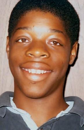
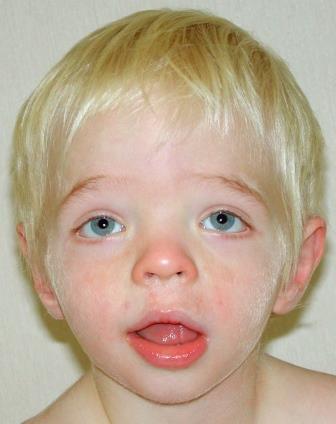
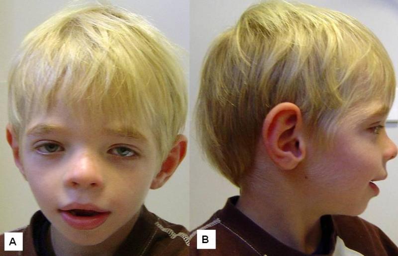
    • Craniofacial features (particularly in an affected older child or adult). Widely spaced eyes with downslanted palpebral fissures; depressed nasal tip with thick alae nasi and broad columella; protruding ears; wide mouth with thick vermilion of the upper and lower lips; coarse face that may further coarsen with age (Figures 1, 2, and 3)
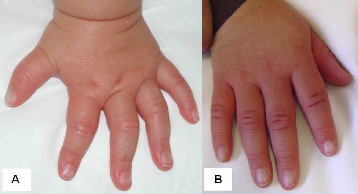
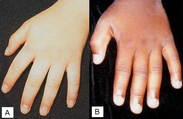
    • Hand findings. Small, soft, fleshy hands; distally tapered fingers with small terminal phalanges and small nails; hyperextensible fingers (Figures 4 and 5)

Females

  • Developmental delay / intellectual disability most often presents as variable degrees of developmental delay with or without intellectual disability. When present, intellectual disability is generally mild to moderate in degree.
  • Neurologic and musculoskeletal findings may manifest variably in heterozygous females, often to a milder degree.
  • Physical characteristics
    • Craniofacial features. Females may have subtle craniofacial features, and some have been reported with features similar to those seen in males.
    • Hand findings. Females may have mildly tapered, soft, fleshy fingers.

Imaging Findings

Radiographic findings that are nonspecific individually or as a pattern but may be helpful when the diagnosis is suspected include metacarpal pseudoepiphyses, poor modeling of the middle phalanges, and tufting of the distal phalanges (metacarpophalangeal profiles do not appear to aid diagnosis) [Hanauer & Young 2002].

Brain MRI. In some individuals, mild cerebral atrophy, hypoplasia of the corpus callosum, periventricular white matter changes in the parietal and frontal lobes, and/or constriction of the foramen magnum may be observed [Tos et al 2015, Upadia et al 2017, Miyata et al 2018].

Family History

Family history is consistent with X-linked inheritance (e.g., no male-to-male transmission). Absence of a known family history does not preclude the diagnosis.

Establishing the Diagnosis

Male proband. The diagnosis of RPS6KA3-ID is established in a male proband with suggestive findings and a hemizygous pathogenic (or likely pathogenic) variant in RPS6KA3 identified by molecular genetic testing (see Table 1).

Female proband. The diagnosis of RPS6KA3-ID is usually established in a female proband with suggestive findings and a heterozygous pathogenic (or likely pathogenic) variant in RPS6KA3 identified by molecular genetic testing (see Table 1).

Note: (1) Per ACMG/AMP variant interpretation guidelines, the terms "pathogenic variants" and "likely pathogenic variants" are synonymous in a clinical setting, meaning that both are considered diagnostic and both can be used for clinical decision making [Richards et al 2015]. Reference to "pathogenic variants" in this section is understood to include any likely pathogenic variants. (2) Identification of a hemizygous or heterozygous RPS6KA3 variant of uncertain significance does not establish or rule out the diagnosis.

Genetic mechanisms other than monoallelic pathogenic variants in RPS6KA3 have not been reported in affected females, with the exception of a female who had a balanced X;autosome translocation that directly disrupted one copy of RPS6KA3, resulting in preferential inactivation of the normal X chromosome [Yamoto et al 2020].

Molecular genetic testing approaches can include a combination of gene-targeted testing (single-gene testing, multigene panel) and comprehensive genomic testing (chromosomal microarray analysis, exome sequencing, genome sequencing) depending on the phenotype.

Gene-targeted testing requires that the clinician determine which gene(s) are likely involved, whereas genomic testing does not. Individuals with the distinctive findings described in Suggestive Findings are likely to be diagnosed using gene-targeted testing (see Option 1), whereas those in whom the diagnosis of RPS6KA3-ID spectrum has not been considered are more likely to be diagnosed using genomic testing (see Option 2).

Option 1

An X-linked intellectual disability, epilepsy, or spastic paraplegia multigene panel that includes RPS6KA3 and other genes of interest (see Differential Diagnosis) is most likely to identify the genetic cause of the condition while limiting identification of variants of uncertain significance and pathogenic variants in genes that do not explain the underlying phenotype. Note: (1) The genes included in the panel and the diagnostic sensitivity of the testing used for each gene vary by laboratory and are likely to change over time. (2) Some multigene panels may include genes not associated with the condition discussed in this GeneReview. (3) In some laboratories, panel options may include a custom laboratory-designed panel and/or custom phenotype-focused exome analysis that includes genes specified by the clinician. (4) Methods used in a panel may include sequence analysis, deletion/duplication analysis, and/or other non-sequencing-based tests.

For an introduction to multigene panels click here. More detailed information for clinicians ordering genetic tests can be found here.

Option 2

When the diagnosis of RPS6KA3-ID has not been considered because an individual has developmental delay / intellectual disability without other suggestive phenotypic features or a suggestive family history, comprehensive genomic testing does not require the clinician to determine which gene is likely involved. Exome sequencing is most commonly used; genome sequencing is also possible.

For an introduction to comprehensive genomic testing click here. More detailed information for clinicians ordering genomic testing can be found here.

Table 1.

Molecular Genetic Testing Used in RPS6KA3-Related Intellectual Disability

Gene 1MethodProportion of Pathogenic Variants 2 Detectable by Method
RPS6KA3 Sequence analysis 3, 485%-90% 5
Gene-targeted deletion/duplication analysis 610%-15% 5
1.

See Table A. Genes and Databases for chromosome locus and protein.

2.

See Molecular Genetics for information on variants detected in this gene.

3.

Sequence analysis detects variants that are benign, likely benign, of uncertain significance, likely pathogenic, or pathogenic. Variants may include small intragenic deletions/insertions and missense, nonsense, and splice site variants; typically, exon or whole-gene deletions/duplications are not detected. For issues to consider in interpretation of sequence analysis results, click here.

4.

Schneider et al [2013] identified a deep intronic pathogenic variant that resulted in an aberrant protein.

5.

Data derived from the subscription-based professional view of Human Gene Mutation Database [Stenson et al 2020]

6.

Gene-targeted deletion/duplication analysis detects intragenic deletions or duplications. Methods used may include a range of techniques such as quantitative PCR, long-range PCR, multiplex ligation-dependent probe amplification (MLPA), and a gene-targeted microarray designed to detect single-exon deletions or duplications.

Clinical Characteristics

Clinical Description

At the more severe end of the continuum of RPS6KA3-related intellectual disability (RPS6KA3-ID), clinically described Coffin-Lowry syndrome commonly involves developmental delay, intellectual disability, neurologic manifestations (hypotonia, stimulus-induced drop attacks, spastic paraparesis, and seizures), musculoskeletal manifestations (kyphoscoliosis and pectus deformity), and characteristic craniofacial and hand findings. The milder end of the continuum primarily manifests with neurodevelopmental features and variable but less pronounced multisystem involvement. Some heterozygous females exhibit clinical manifestations that are typically less severe than those seen in males. However, females can have recognizable craniofacial and hand findings that suggest the diagnosis of RPS6KA3-ID.

To date, more than 200 individuals have been identified with a pathogenic variant in RPS6KA3. A precise number is not known due to literature reports of individuals who were diagnosed based on clinical features prior to gene identification. The following description of the phenotypic features associated with RPS6KA3-ID is derived from an admixture of reported individuals whose diagnoses were molecularly confirmed as well as reported individuals who were diagnosed based on clinical findings, radiographic findings, and/or family history without molecular confirmation.

Affected Males

Table 2.

RPS6KA3-Related Intellectual Disability: Frequency of Select Features in Males

FeatureFrequency 1
Development Developmental delay (typically severe)+++ (100%)
Intellectual disability+++
Neurobehavioral/psychiatric +
Neurologic Stimulus-induced drop attacks+ (13%-20%)
Progressive spasticity/paraplegia++
Seizures+ (5%-30%)
Musculoskeletal Kyphoscoliosis or other spine deformity++ (40%-80%)
Pectus carinatum/excavatum+++ (80%)
Cardiovascular Cardiomyopathy+
Valvular abnormalities+
Reduced height +++
Dental issues +
Hearing loss ++ (30%)
Vision issues +
Sleep apnea +

+++ = high frequency; ++ = moderate frequency, + = low frequency

1.

Relative frequencies are provided, with percentages in parentheses where those are available in the literature

Developmental delay (DD) and intellectual disability (ID). Early milestones are variably delayed, with speech typically more severely affected than motor development. Coffin-Lowry syndrome (CLS) is typically characterized by severe-to-profound ID in males, although those with mild disability have been reported [Hanauer & Young 2002, Hunter 2002, Pereira et al 2010]. These individuals may now be better classified as having RPS6KA3-ID. Early developmental assessments may overestimate the ultimate developmental prognosis [Hunter 2002]. Manouvrier-Hanu et al [1999] reported two sibs with an unusually mild presentation associated with a missense variant.

Neurobehavioral/psychiatric manifestations. Males with CLS are often described as generally happy and easygoing, although behavioral issues can occur. These include attention-deficit/hyperactivity disorder [Matsumoto et al 2013], aggressive behavior [Hunter 2002], self-injury, and manifestations of autism spectrum disorder.

Neurologic. Detailed neurologic assessment may be hampered by the presence of severe ID. Findings reported include the following:

  • Stimulus-induced drop attacks (SIDAs) (also called stimulus-induced drop episodes, or SIDEs), which occur when an unexpected tactile or auditory stimulus or excitement triggers a brief electromyographic silence in the lower limbs, resulting in a brief collapse (though no loss of consciousness) [Hahn & Hanauer 2012].
    • In most cases, the transient loss of muscle tone occurs in the paraspinal or quadriceps muscles. Some individuals also experience myoclonic jerks or increased muscle tone due to "hyperekplexia-like" episodes with a startle response [Hahn & Hanauer 2012].
    • Onset occurs between ages four and 17 years with a mean age of 8.6 years [Nakamura et al 2005].
    • SIDAs were reported in 20% (34/170) of individuals in the CLS Foundation database [Stephenson et al 2005].
    • The episodes may become debilitating due to injury from falls [Nelson & Hahn 2003, Hahn & Hanauer 2012].
    • See Nelson & Hahn [2003] for a video illustration of SIDAs.
  • Progressive spasticity/paraplegia with loss of the ability to walk is ascribed to both calcification of the ligamenta flava and stenosis of the spinal canal [Hunter 2002, Welborn et al 2018]. Loss of strength and muscle mass and both decreased and increased deep tendon reflexes have been observed.
  • Seizures. True epileptic seizures are thought to affect at least 5% of individuals with CLS [Stephenson et al 2005, Gschwind et al 2015]. Some have described a risk of 5%-30% [Hahn & Hanauer 2012]. Seizures require differentiation from SIDAs or other atypical movements that can occur in persons with RPS6KA3-ID but are not epileptic in origin [Stephenson et al 2005]. Hahn & Hanauer [2012] noted that seizures in persons with CLS often last longer than SIDAs, have a focal onset, and can be associated with clonic movements and tonic posturing.
  • Feeding difficulties due to hypotonia may present early in life.
  • Neuroimaging studies have not demonstrated a consistent pattern of brain abnormalities, but the following findings have been reported [Tos et al 2015, Upadia et al 2017]:
    • Abnormalities of the corpus callosum, including thinning and agenesis [Wang et al 2006]
    • Multiple focal frontal hypodensities visible on MRI. Hypodensities attributed to focal areas of cerebrospinal fluid were reported in three affected sibs by Wang et al [2006]; the sibs also showed thinning of the corpus callosum, vermian hypoplasia, and mild ventricular asymmetry. The authors concluded that the degree of ID correlated with the severity of the MRI findings.
    • Periventricular white matter abnormalities/cysts [Miyata et al 2018]
    • Constricted foramen magnum (decreased diameter)
    • Reduced gray and white matter volume without evidence of ventriculomegaly ex vacuo on quantitative MRI in affected males and females, suggesting an early neurodevelopmental abnormality such as reduced cellular proliferation [Kesler et al 2007]

Musculoskeletal

  • Progressive kyphoscoliosis is one of the most difficult aspects of the long-term care of individuals with CLS. At least 40% of affected males have been reported to have progressive kyphoscoliosis [Hunter 2002]. The rates were higher in a series reported from an orthopedic referral clinic [Herrera-Soto et al 2007]. When other types of abnormalities such as thoracic lordosis and degenerative disc disease are included, the rate of spinal abnormalities in persons with CLS approaches 80% [Welborn et al 2018]. Calcifications and/or hypertrophy of the ligamentum flavum have been noted in several reports, and other findings may include irregular vertebral end plates, anterior wedging, and narrowing of the intervertebral spaces [Welborn et al 2018].
    The severity of the spinal deformity often worsens significantly over time and can lead to severe complications:
    • Cardiorespiratory compromise caused by kyphoscoliosis may contribute to premature death.
    • Neurologic compromise can also occur, and rapidly progressive kyphosis with acute paralysis has been reported in several instances. Welborn et al [2018] evaluated eight males ranging in age from 13 to 22 years and found that four had marked calcifications and two had marked hypertrophy of the ligamentum flavum in the cervical region. They also noted that 100% of those in their series with calcifications of the ligamentum flavum had neurologic abnormalities, including marked lower extremity weakness and acute quadriparesis. Furthermore, they found that the calcifications could be visualized on CT scan many years before the development of neurologic abnormalities, suggesting that ligamentum flavum calcifications of this type may be useful indicators for closer monitoring and potentially earlier surgical intervention.
  • Pectus carinatum and/or excavatum are frequently seen.
  • Pes planus may occur [Welborn et al 2018].
  • Other minor skeletal changes that may be seen on radiographs are of no clinical consequence.

Cardiovascular. Approximately 14% of affected males have cardiovascular disorders [Hunter 2002, Martinez et al 2011, Yoshida et al 2015, Wakami et al 2022]. This percentage may be an underestimate, as many individuals with CLS have not had thorough initial or ongoing cardiac assessment. Reports have included abnormalities of the mitral, tricuspid, and aortic valves, short chordae, cardiomyopathy (with endocardial fibroelastosis in one individual), unexplained congestive heart failure, and dilatation of the aorta and of the pulmonary artery. Facher et al [2004] reported a 14-year-old male with restrictive cardiomyopathy. Martinez et al [2011] reported a male with CLS who had left ventricular non-compaction cardiomyopathy with a restrictive pattern. Corrective surgery for mitral and triscupid insufficiency has been described in two males at ages 14 and 18 years, respectively [Yoshida et al 2015, Wakami et al 2022]. Cardiac anomalies may contribute to premature death. There has not been a systematic review of cardiovascular disorders in individuals with CLS.

Growth. Prenatal growth is usually normal; reduced growth usually occurs early in the postnatal period [Touraine et al 2002]. Males generally fall below the third centile in height but are expected to track a curve. Height in adult males has been reported to range between 115 and 158 cm, with an average of 143 cm [Hanauer & Young 2002]. Kyphoscoliosis may exacerbate the reduction in stature [Touraine et al 2002]. The safety and efficacy of growth hormone for the treatment of short stature in persons with CLS have not been studied, and it has been suggested that this could aggravate skeletal deformities and calcifications of the ligamentum flavum [Lv et al 2019].

While microcephaly is common, many individuals with CLS have a normal head circumference. Short stature, hypotonia, and decreased activity may lead to increased risk for obesity.

Dental. Dental anomalies commonly include small teeth, malpositioning, open bite, hypodontia of secondary teeth, advanced or delayed eruption of primary teeth, and premature loss [Hunter 2002, Igari et al 2006, Norderyd & Aronsson 2012]. The palate is high. With age, the retrognathia in the younger child tends to be replaced by prognathism.

Hearing loss is reported in as many as 30% of individuals [Pereira et al 2010]. Hunter [2002] reported hearing loss in 14 of 89 affected males.

  • An audiogram may reveal sensorineural hearing loss. Mixed hearing loss has also been described [Hunter 2002].
  • Malformation of the labyrinth has been reported [Hunter 2002], as has late onset of hearing loss.
  • Clustering of hearing loss within families may occur.

Vision. Significant visual issues appear to be uncommon, although cataract, retinal pigment atrophy, and optic atrophy have been reported, and the incidence of chronic eyelid irritation (blepharitis) may be increased [Hunter 2002].

Respiratory. Obstructive sleep apnea may occur. Tracheostomy was reported to improve both obstructive sleep apnea and SIDAs in an individual reported by Imataka et al [2016].

Persons with CLS may have issues with intubation and/or ventilation that require careful consideration of the strategy for airway management during anesthesia [Hirakawa et al 2017].

Restrictive lung disease may occur due to kyphoscoliosis; this can be severe and go unrecognized [Venter et al 2019].

Other. Findings reported in single individuals include rectal prolapse, jejunal diverticuli, colonic diverticuli with reduced ganglion cells, popliteal ganglion, pyloric stenosis, unilateral renal agenesis, anteriorly placed anus, increased facial pigment, and enlarged trachea [Hunter 2002]. A family was reported in which multiple individuals developed type II diabetes [Touma Boulos et al 2021].

Prognosis. A number of adult males with CLS have been described. One recently reported individual is alive at age 48 years [Di Stazio et al 2021]. Since many adults with disabilities have not undergone advanced genetic testing, it is likely that adults with RPS6KA3-ID are underrecognized and underreported.

Life span is reduced in some males with CLS. Of individuals reported in the literature, death occurred in 13.5% of males at a mean age of 20.5 years (range: 13-34) [Hunter 2002].

  • Complicating factors have included cardiac anomalies, panacinar emphysema, respiratory complications, progressive kyphoscoliosis, and seizure-associated aspiration.
  • Coffin [2003] reported that one of his original patients died at age 18.8 years of pneumonia superimposed on chronic lung and heart disease, and a second individual died at age 18 years of acute food aspiration.
  • Hanauer & Young [2002] noted that several males died following complications related to general anesthesia.
  • The authors are aware of an individual with CLS who had life-threatening central and obstructive sleep apnea, and of another male who had a history of chronic obstructive and central sleep apnea who died from respiratory complications after surgery for jaw advancement.
  • One affected male died of Hodgkin disease [Hunter 2002].

Heterozygous Females

Heterozygous females often manifest markedly variable clinical manifestations of RPS6KA3-ID, such as mild facial coarsening, tapered fingers, short stature, and varied degrees of ID. Females with facial, hand, and skeletal findings typical of those seen in hemizygous males have been reported [Fryssira et al 2002, Hunter 2002, Jurkiewicz et al 2010, Rojnueangnit et al 2014]. Some heterozygous females have typical development and intellectual ability and lack other systemic findings associated with RPS6KA3-ID.

DD and ID. Affected females tend to have ID in the mild-to-moderate range.

Neurobehavioral/psychiatric manifestations. The rate of psychiatric illness may be higher than that in the general population. Six (8.8%) of 68 women (22 females with RPS6KA3-ID, 38 unaffected heterozygotes, and 8 "affected" sisters) have had psychiatric diagnoses, including schizophrenia, bipolar disease, and "psychosis" (reviewed in Hunter [2002]). One of two women reported by Micheli et al [2007] had a "psychosis," and one of two affected sisters was reported by Wang et al [2006] to have schizophrenia. Pervasive developmental disorder has been described [Matsumoto et al 2013]. Compulsive eyebrow-pulling behavior was reported in one female [Gürsoy et al 2022].

Neurologic. Females with typical SIDAs have been reported [Jurkiewicz et al 2010, Arslan et al 2014, Rojnueangnit et al 2014].

Musculoskeletal. At least 32% of females have been reported to have progressive kyphoscoliosis [Hunter 2002]. Rojnueangnit et al [2014] reported a female who was diagnosed with both scoliosis and spondylolisthesis at age 10 years and required surgical fusion from L4 to S1 by age 11 years due to the severity and progressive nature of her spinal issues.

Cardiovascular. Approximately 5% of affected females have cardiovascular disorders [Hunter 2002]. Cong et al [2022] described a family in which three females with a pathogenic variant in RPS6KA3 all had mild mitral and tricuspid regurgitation. However, it should be noted that two of these individuals also had a distal chromosome 22q11 deletion, which may have contributed to their phenotypes.

Growth. Stature may be reduced or fall within the typical range.

Dental. Females may have dental manifestations, including hypodontia [Jurkiewicz et al 2010, Yamoto et al 2020, Song et al 2022].

Hearing loss. Hunter [2002] reported hearing loss in one of 22 affected females.

Vision. Specific data are not available regarding the frequency or types of vision issues observed in females with RPS6KA3-ID.

Respiratory. Sleep apnea may occur.

Other. Idiopathic hypercalcemia requiring bisphosphonate therapy during the second year of life was described in a female with RPS6KA3-ID [Tise et al 2022]. Genitourinary tract anomalies including uterine prolapse, bicornuate uterus, and duplicationof the renal collecting system have also been reported [Hunter 2002, Tise et al 2022]. Central precocious puberty with advanced bone age was described in an affected female [Song et al 2022].

Prognosis. Data are lacking as to whether life span in females with RPS6KA3-ID is impacted. Hunter [2002] noted that one affected female had died at age 48.
Genotype-Phenotype Correlations

Although no strong correlation exists between phenotype and location or type of RPS6KA3 pathogenic variant, individuals with certain missense pathogenic variants may tend to have milder disease expression [Delaunoy et al 2001].

Nakamura et al [2005] suggested that truncating variants, either in or upstream from the N-terminal kinase domain, may cause a particular susceptibility to stimulus-induced drop attacks (SIDAs). However, the finding of an affected female with SIDAs who had a heterozygous pathogenic variant in the region encoding the C-terminal kinase domain of the protein would argue against this correlation [Rojnueangnit et al 2014].

Nomenclature

Early authors referred to Coffin syndrome until it was recognized that the individuals reported by Lowry et al [1971] had the same syndrome.

Some early texts and papers confused Coffin-Siris syndrome and Coffin-Lowry syndrome.

RPS6KA3-related X-linked nonsyndromic intellectual disability – a phenotype at the milder end of the RPS6KA3-ID spectrum – was originally referred to as MRX19 (OMIM 300844).

The title of this GeneReview, "RPS6KA3-related intellectual disability," is based on the dyadic naming approach proposed by Biesecker et al [2021] to delineate mendelian genetic disorders.

Prevalence

No estimate of the prevalence of CLS has been published. Based on the authors' experience, a rate of 1:40,000-50,000 may be reasonable – although it may underestimate the actual prevalence.

Differential Diagnosis

Individuals with phenotypic findings suggestive of Coffin-Lowry syndrome (the more severe end of the RPS6KA3-related intellectual disability phenotypic continuum). See Table 3.

Table 3.

Disorders of Interest in the Differential Diagnosis of Coffin-Lowry Syndrome

Gene / Genetic MechanismDisorderMOIClinical Characteristics / Comment
ATRX Alpha-thalassemia X-linked intellectual disability syndrome XLDistinctive craniofacial features, genital anomalies, hypotonia, mild-to-profound DD/ID. While all affected persons have a normal 46,XY karyotype, genital anomalies comprise a range from hypospadias & undescended testicles, to severe hypospadias & ambiguous genitalia, to normal-appearing female external genitalia. Alpha-thalassemia (~75% of affected persons) is mild & typically does not require treatment.
MED12 FG syndrome type 1 (FGS1) (See MED12-Related Disorders.)XLFGS1 shares w/CLS broad forehead, widely spaced eyes w/downslanted palpebral fissures, thick vermilion of the lower lip, kyphoscoliosis, pectus excavatum, & characteristic behaviors. Unlike CLS, FGS1 is assoc w/disproportionate macrocephaly; constipation that may be assoc w/anal anomalies; broad thumbs & halluces; prominent fingertip pads; & small, rounded, cupped ears that often have overfolded superior helix. Hypotonia often evolves into joint restriction. Partial absence of corpus callosum & fused mammillary bodies are relatively common.
PHF6

Borjeson-Forssman-Lehmann syndrome (BFLS) (OMIM 301900)

XLSevere ID, hand findings similar to those of CLS, short nose w/anteverted nares that may be small w/thick septum, & kyphoscoliosis. Additional findings are large, prominent ears & visual issues. Affected males have extreme hypogonadism & tend to have marked gynecomastia. Females may show partial expression of syndrome.
TCF4 1 Pitt-Hopkins syndrome See footnote 2.Distinctive facial features that become more apparent w/age, significant DD/ID, & episodic hyperventilation &/or breath-holding while awake (~50% of affected persons). Other common findings are behavioral issues, hand stereotypies, seizures, constipation, & severe myopia.
7q11.23 contiguous gene deletion of WBSCR Williams syndrome AD 3In addition to facial findings that may resemble those in CLS, Williams syndrome is assoc w/cardiovascular disease (elastin arteriopathy, peripheral pulmonary stenosis, supravalvar aortic stenosis, hypertension), connective tissue abnormalities, ID (usually mild), a specific cognitive profile, unique personality characteristics, growth abnormalities, & endocrine abnormalities (hypercalcemia, hypercalciuria, hypothyroidism, & early puberty). Feeding difficulties often lead to failure to thrive in infancy.

AD = autosomal dominant; CLS = Coffin-Lowry syndrome; DD = developmental delay; ID = intellectual disability; MOI = mode of inheritance; WBSCR = Williams-Beuren syndrome critical region; XL = X-linked

1.

Pitt-Hopkins syndrome is caused by haploinsufficiency of TCF4 resulting from either a pathogenic variant in TCF4 or a deletion of the chromosome region in which TCF4 is located (18q21.2).

2.

In most affected individuals, Pitt-Hopkins syndrome results from a de novo pathogenic variant or deletion.

3.

Most cases are de novo occurrences, but occasionally parent-to-child transmission is observed.

Individuals with developmental delay / intellectual disability without other suggestive phenotypic features. At the milder end of the RPS6KA3-ID phenotypic continuum, neurodevelopmental features and variable multisystem involvement are not sufficient to diagnose RPS6KA3-ID clinically; thus, many other disorders with intellectual disability should be considered in the differential diagnosis. See OMIM Autosomal Dominant, Autosomal Recessive, and Syndromic X-Linked Intellectual Developmental Disorder Phenotypic Series.

Management

No clinical practice guidelines for RPS6KA3-related intellectual disability (RPS6KA3-ID) have been published.

Evaluations Following Initial Diagnosis

To establish the extent of disease and needs in an individual diagnosed with RPS6KA3-ID, the evaluations summarized in Table 4 (if not performed as part of the evaluation that led to the diagnosis) are recommended.

Table 4.

Recommended Evaluations Following Initial Diagnosis in Individuals with RPS6KA3-Related Intellectual Disability

System/ConcernEvaluationComment
Development Developmental assessment
  • To incl motor, adaptive, cognitive, & speech-language eval
  • Eval for early intervention / special education
Neurobehavioral/
Psychiatric
Neuropsychiatric evalFor persons age >12 mos: screening for behavior concerns incl sleep disturbances, ADHD, &/or findings suggestive of ASD
Neurologic Neurologic eval
  • To assess for SIDAs, changes in gait or in bowel or bladder function, & seizures or movement disorder
  • Consider EEG if seizures are a concern.
Musculoskeletal Orthopedics / physical medicine & rehab / PT & OT evalTo incl assessment of:
  • Gross motor & fine motor skills
  • Kyphoscoliosis
  • Pectus deformity
  • Contractures
  • Mobility, ADL, & need for adaptive devices
  • Need for PT (to improve gross motor skills) &/or OT (to improve fine motor skills)
Cardiovascular Cardiology evalTo incl assessment of:
  • Cardiomyopathy
  • Valve dysfunction or other abnormalities
Growth Measurement of height, weight, & head circumferenceWith attention to pattern of height growth under expectation of ↓ stature but normal growth velocity
Dental Dental evalTo incl assessment of:
  • Small or malpositioned teeth
  • Advanced or delayed eruption of primary teeth
  • Hypodontia of secondary teeth
  • Premature tooth loss
Hearing Audiologic evalAssess for sensorineural hearing loss.
Eyes Ophthalmologic evalTo assess for ↓ vision, abnormal ocular movement, best corrected visual acuity, refractive errors, strabismus, blepharitis, & more complex findings (e.g., cataract, retinal pigment atrophy, optic atrophy) that may require referral for subspecialty care &/or low vision services
Respiratory Sleep medicine eval
  • To assess for obstructive sleep apnea
  • Refer to pulmonologist to evaluate for restrictive lung disease if severe kyphoscoliosis or concerning respiratory symptoms are present.
Genetic counseling By genetics professionals 1To inform affected persons & their families re nature, MOI, & implications of RPS6KA3-ID to facilitate medical & personal decision making
Family support
& resources
Assess need for:

ADHD = attention-deficit/hyperactivity disorder; ADL = activities of daily living; ASD = autism spectrum disorder; MOI = mode of inheritance; OT = occupational therapy; PT = physical therapy; RPS6KA3-ID = RPS6KA3-related intellectual disability

1.

Medical geneticist, certified genetic counselor, certified advanced genetic nurse

Treatment of Manifestations

There is no cure for RPS6KA3-ID.

Supportive care to improve quality of life, maximize function, and reduce complications is recommended. This ideally involves multidisciplinary care by specialists in relevant fields (see Table 5).

Table 5.

Treatment of Manifestations in Individuals with RPS6KA3-Related Intellectual Disability

Manifestation/ConcernTreatmentConsiderations/Other
Developmental delay /
Intellectual disability /
Neurobehavioral/
psychiatric issues
See Developmental Delay / Intellectual Disability Management Issues.Risperidone may be of benefit to persons who display destructive or self-injurious behavior [Valdovinos et al 2002].
Stimulus-induced drop attacks (SIDAs)
  • A trial of medication & efforts to optimize dosage may be considerations. 1 Benzodiazepines have often been used as first-line treatment. 2
  • Awareness of SIDAs should allow early intervention to minimize occurrence of triggering stimuli & provide protection from falls.
  • If attacks occur w/great frequency a protective helmet may be indicated & use of a wheelchair may be required to prevent falling & injury.
  • Various medications have been used in an attempt to manage SIDAs; these incl benzodiazepines, ASMs, SSRIs, & tricyclic antidepressants.
  • While most treatments have not resulted in significant long-term control of SIDAs, improvements in selected persons have been seen w/clonazepam, 3 unspecified benzodiazepines, 4 sodium oxybate, 5 fluoxetine, & clomipramine. 6
  • Valproic acid led to improvement in 1 persons, but this & other conventional ASMs have generally been ineffective. 2
  • Improvement of SIDAs occurred in a male after treatment of obstructive sleep apnea w/tracheostomy. 7
Spasticity/
Paraplegia
Orthopedics / physical medicine & rehab / PT & OT incl stretching to help avoid contractures & falls
  • Consider need for positioning & mobility devices, disability parking placard.
Seizures Standardized treatment w/ASM by experienced neurologist
  • Many ASMs may be effective; none has been demonstrated effective specifically for this disorder.
  • Education of parents/caregivers 8
Kyphoscoliosis Per orthopedistBracing or spine surgery may be indicated to prevent pulmonary volume restriction & neurologic compromise.
Pectus carinatum/
excavatum
Per orthopedist &/or general surgeonSevere pectus excavatum may require surgery to improve thoracic volume.
Cardiomyopathy, valve dysfunction, or other cardiovascular abnormalities Per cardiologistPhysical activity may be limited as recommended by cardiologist.
Growth issues Per nutritionist &/or feeding therapist
  • Ensure appropriate nutritional & feeing support in hypotonic infants w/feeding difficulty.
  • Abnormal growth velocity & obesity should be assessed & treated in standard manner.
  • There are no data regarding safety or efficacy of growth hormone for treatment of growth deficiency in this disorder.
Dental issues Per dentistStandard dental care
Hearing Hearing aids may be helpful per otolaryngologist.Community hearing services through early intervention or school district
Eyes Per ophthalmologistRefractive errors, strabismus
Per ophthalmic subspecialistMore complex findings (e.g., cataract, retinal pigment atrophy, optic atrophy)
Per low vision services
  • Children: through early intervention programs &/or school district
  • Adults: low vision clinic &/or community vision services / OT / mobility services
Respiratory Per sleep specialistCPAP or surgery may be helpful in mgmt of obstructive sleep apnea.
Per otolaryngologist &/or anesthesiologistAffected persons may have issues w/intubation &/or ventilation, making it important to address any potential concerns regarding respiratory mgmt prior to surgery or other procedures requiring anesthesia or sedation.
Family/Community
  • Ensure appropriate social work involvement to connect families w/local resources, respite, & support.
  • Coordinate care to manage multiple subspecialty appointments, equipment, medications, & supplies.
  • Ongoing assessment of need for palliative care involvement &/or home nursing
  • Consider involvement in adaptive sports or Special Olympics.

ASM = anti-seizure medication; CPAP = continuous positive airway pressure; OT = occupational therapy; PT = physical therapy; SIDAs = stimulus-induced drop attacks; SSRIs = selective serotonin reuptake inhibitors

1.
2.
3.
4.
5.
6.
7.
8.

Education of parents/caregivers regarding common seizure presentations is appropriate. For information on non-medical interventions and coping strategies for children diagnosed with epilepsy, see Epilepsy Foundation Toolbox.

Developmental Delay / Intellectual Disability Management Issues

The following information represents typical management recommendations for individuals with developmental delay / intellectual disability in the United States; standard recommendations may vary from country to country.

Ages 0-3 years. Referral to an early intervention program is recommended for access to occupational, physical, speech, and feeding therapy as well as infant mental health services, special educators, and sensory impairment specialists. In the US, early intervention is a federally funded program available in all states that provides in-home services to target individual therapy needs.

Ages 3-5 years. In the US, developmental preschool through the local public school district is recommended. Before placement, an evaluation is made to determine needed services and therapies and an individualized education plan (IEP) is developed for those who qualify based on established motor, language, social, or cognitive delay. The early intervention program typically assists with this transition. Developmental preschool is center based; for children too medically unstable to attend, home-based services are provided.

All ages. Consultation with a developmental pediatrician is recommended to ensure the involvement of appropriate community, state, and educational agencies (US) and to support parents in maximizing quality of life. Some issues to consider:

  • IEP services:
    • An IEP provides specially designed instruction and related services to children who qualify.
    • IEP services will be reviewed annually to determine whether any changes are needed.
    • Special education law requires that children participating in an IEP be in the least restrictive environment feasible at school and included in general education as much as possible, when and where appropriate.
    • Vision and hearing consultants should be a part of the child's IEP team to support access to academic material.
    • PT, OT, and speech services will be provided in the IEP to the extent that the need affects the child's access to academic material. Beyond that, private supportive therapies based on the affected individual's needs may be considered. Specific recommendations regarding type of therapy can be made by a developmental pediatrician.
    • As a child enters the teen years, a transition plan should be discussed and incorporated in the IEP. For those receiving IEP services, the public school district is required to provide services until age 21.
  • A 504 plan (Section 504: a US federal statute that prohibits discrimination based on disability) can be considered for those who require accommodations or modifications such as front-of-class seating, assistive technology devices, classroom scribes, extra time between classes, modified assignments, and enlarged text.
  • Developmental Disabilities Administration (DDA) enrollment is recommended. DDA is a US public agency that provides services and support to qualified individuals. Eligibility differs by state but is typically determined by diagnosis and/or associated cognitive/adaptive disabilities.
  • Families with limited income and resources may also qualify for supplemental security income (SSI) for their child with a disability.

Motor Dysfunction

Gross motor dysfunction

  • Physical therapy is recommended to maximize mobility and to reduce the risk for later-onset orthopedic complications (e.g., contractures, scoliosis, hip dislocation).
  • Consider use of durable medical equipment and positioning devices as needed (e.g., wheelchairs, walkers, bath chairs, orthotics, adaptive strollers).
  • For muscle tone abnormalities including hypertonia or dystonia, consider involving appropriate specialists to aid in management of baclofen, tizanidine, Botox®, anti-parkinsonian medications, or orthopedic procedures.

Fine motor dysfunction. Occupational therapy is recommended for difficulty with fine motor skills that affect adaptive function such as feeding, grooming, dressing, and writing.

Oral motor dysfunction should be assessed at each visit and clinical feeding evaluations and/or radiographic swallowing studies should be obtained for choking/gagging during feeds, poor weight gain, frequent respiratory illnesses, or feeding refusal that is not otherwise explained. Assuming that the child is safe to eat by mouth, feeding therapy (typically from an occupational or speech therapist) is recommended to help improve coordination or sensory-related feeding issues. Feeds can be thickened or chilled for safety. When feeding dysfunction is severe, an NG-tube or G-tube may be necessary.

Communication issues. Consider evaluation for alternative means of communication (e.g., augmentative and alternative communication [AAC]) for individuals who have expressive language difficulties. An AAC evaluation can be completed by a speech-language pathologist who has expertise in the area. The evaluation will consider cognitive abilities and sensory impairments to determine the most appropriate form of communication. AAC devices can range from low-tech, such as picture exchange communication, to high-tech, such as voice-generating devices. Contrary to popular belief, AAC devices do not hinder verbal development of speech, but rather support optimal speech and language development.

Neurobehavioral/Psychiatric Concerns

Children may qualify for and benefit from interventions used in treatment of autism spectrum disorder, including applied behavior analysis (ABA). ABA therapy is targeted to the individual child's behavioral, social, and adaptive strengths and weaknesses and typically performed one on one with a board-certified behavior analyst.

Consultation with a developmental pediatrician may be helpful in guiding parents through appropriate behavior management strategies or providing prescription medications, such as medication used to treat attention-deficit/hyperactivity disorder, when necessary.

Concerns about serious aggressive or destructive behavior can be addressed by a pediatric psychiatrist.

Surveillance

To monitor existing manifestations, the individual's response to supportive care, and the emergence of new manifestations, the evaluations summarized in Table 6 are recommended.

Table 6.

Recommended Surveillance for Individuals with RPS6KA3-Related Intellectual Disability

System/ConcernEvaluationFrequency
Development Monitor developmental progress & educational needs.At each visit
Neurobehavioral/
Psychiatric
Behavioral assessment for ADHD, aggressive behavior, & ASD
Neurologic Monitor those w/SIDAs, spasticity, or seizures.Per treating neurologist
Assess for new manifestations of SIDAs & seizures.At each visit
Assess for new/worsening signs/symptoms of spinal canal narrowing, incl changes in tone, gait, & bowel/bladder habits, expression of pain, & focal neurologic changes such as clonus or abnormal tendon reflexes.
  • At each visit
  • Note: Persons w/calcifications of ligamentum flavum, particularly in cervical region, may require more careful surveillance. 1
Musculoskeletal Physical medicine, OT/PT assessment of mobility, self-help skillsAt each visit
Monitor those w/kyphoscoliosis or pectus deformity for progression.
  • Per treating orthopedist
  • Note: Skeletal maturity may be delayed, requiring longer than anticipated monitoring for progression. 1
Monitor for calcifications or hypertrophy of ligamentum flavum.
  • Per treating orthopedist
  • Note: Persons w/calcifications of ligamentum flavum, particularly in cervical region, may require more careful surveillance. 1
Cardiovascular Monitor those w/cardiomyopathy, valve dysfunction, or other abnormalities.Per treating cardiologist
Assess for new onset of cardiomyopathy.Even if initial echocardiogram is normal, it should be repeated every 5-10 yrs in light of uncertainty as to incidence & range in age of onset of cardiomyopathy. 2
Growth Monitor for development of obesity.At each visit
Dental Monitor those w/dental abnormalities; otherwise, routine eval as in general population but w/particular attention to ↑ risk of tooth loss.Per treating dentist
Hearing Monitor those w/hearing loss.Per treating otolaryngologist &/or audiologist
Ophthalmologic involvement Monitor those w/refractive errors, strabismus, blepharitis, & more complex findings (e.g., cataract, retinal pigment atrophy, optic atrophy).Per treating ophthalmologist(s)
Low vision servicesPer treating clinicians
Respiratory Monitor those w/sleep apnea.
Family/Community Assess family need for social work support (e.g., palliative/respite care, home nursing, other local resources), care coordination, or follow-up genetic counseling if new questions arise (e.g., family planning).At each visit

ADHD = attention-deficit/hyperactivity disorder; ASD = autism spectrum disorder; OT = occupational therapy; PT = physical therapy

1.
2.

Note: A table containing suggested guidelines for follow up of individuals with CLS is provided in Hunter [2010].

Agents/Circumstances to Avoid

Care should be taken to avoid specific stimuli that are known to trigger SIDAs in a given individual. Physical activity may be limited due to valvular disease as recommended by a cardiologist.

Evaluation of Relatives at Risk

See Genetic Counseling for issues related to testing of at-risk relatives for genetic counseling purposes.

Therapies Under Investigation

Search ClinicalTrials.gov in the US and EU Clinical Trials Register in Europe for access to information on clinical studies for a wide range of diseases and conditions. Note: There may not be clinical trials for this disorder.

Genetic Counseling

Genetic counseling is the process of providing individuals and families with information on the nature, mode(s) of inheritance, and implications of genetic disorders to help them make informed medical and personal decisions. The following section deals with genetic risk assessment and the use of family history and genetic testing to clarify genetic status for family members; it is not meant to address all personal, cultural, or ethical issues that may arise or to substitute for consultation with a genetics professional. —ED.

Mode of Inheritance

RPS6KA3-related intellectual disability (RPS6KA3-ID) is inherited in an X-linked manner.

Risk to Family Members

Parents of a male proband

  • The father of an affected male will not have the disorder nor will he be hemizygous for the RPS6KA3 pathogenic variant; therefore, he does not require further evaluation/testing.
  • In a family with more than one affected individual, the mother of an affected male is an obligate heterozygote. Note: If a woman has more than one affected child and no other affected relatives and if the RPS6KA3 pathogenic variant cannot be detected in her leukocyte DNA, she most likely has germline mosaicism (maternal germline mosaicism has been observed in CLS [Jacquot et al 1998, Horn et al 2001]).
  • If a male is the only affected family member (i.e., a simplex case), the mother may be a heterozygote, the affected male may have a de novo RPS6KA3 pathogenic variant (in which case the mother is not a heterozygote), or the mother may have somatic/germline mosaicism. Roughly two thirds of individuals with CLS have the disorder as the result of a de novo pathogenic variant [Pereira et al 2010]. Similar data are lacking for persons with clinical manifestations at the milder end of the RPS6KA3-ID continuum.
  • Molecular genetic testing of the mother is recommended to confirm her genetic status and to allow reliable recurrence risk assessment.
    Note: Testing of maternal leukocyte DNA may not detect all instances of somatic mosaicism and will not detect a pathogenic variant that is present in the germ cells only.
  • Detailed evaluation of the parents and review of the extended family history may help distinguish male probands with a de novo pathogenic variant from those with an inherited pathogenic variant. Molecular genetic testing of the mother can typically determine if the pathogenic variant was inherited.

Parents of a female proband

  • A female proband may have inherited the RPS6KA3 pathogenic variant from her mother (or possibly her father) or the pathogenic variant may be de novo.
  • Molecular genetic testing of the mother (and possibly the father, or subsequently the father) is recommended to confirm parental genetic status and to allow reliable recurrence risk assessment.
    Note: Testing of parental leukocyte DNA may not detect all instances of somatic mosaicism and will not detect a pathogenic variant that is present in the germ cells only.

Sibs of a male proband. The risk to the sibs of a male proband depends on the genetic status of the mother:

  • If the mother of the proband has an RPS6KA3 pathogenic variant, the chance of the mother transmitting it in each pregnancy is 50%:
    • Males who inherit the pathogenic variant will be affected;
    • Females who inherit the pathogenic variant will be heterozygous and at high risk to exhibit at least some manifestations of the disorder (see Clinical Description, Heterozygous Females). Heterozygous females may show random X-chromosome inactivation or they may show mild-to-moderate skewing of X-chromosome inactivation that does not correlate with IQ [Simensen et al 2002].
      Note: As expected with random X-chromosome inactivation, a mildly affected mother may have a severely affected daughter.
  • If the proband represents a simplex case (i.e., a single occurrence in a family) and if the RPS6KA3 pathogenic variant cannot be detected in the leukocyte DNA of the mother, sibs are still at increased risk of inheriting an RPS6KA3 pathogenic variant because of the possibility of maternal germline mosaicism (maternal germline mosaicism has been observed in CLS [Jacquot et al 1998, Horn et al 2001]).

Sibs of a female proband. The risk to the sibs of a female proband depends on the genetic status of the parents:

  • If the mother of the proband has an RPS6KA3 pathogenic variant, the chance of transmitting it in each pregnancy is 50% (see Sibs of a male proband).
  • If the proband represents a simplex case (i.e., a single occurrence in a family) and if the RPS6KA3 pathogenic variant cannot be detected in the leukocyte DNA of the mother, sibs are still at increased risk of inheriting an RPS6KA3 pathogenic variant because of the possibility of parental germline mosaicism (maternal germline mosaicism has been observed in CLS [Jacquot et al 1998, Horn et al 2001]).

Offspring of a male proband

  • Males with RPS6KA3-ID transmit the RPS6KA3 pathogenic variant to all of their daughters and none of their sons.
  • Severely affected males typically do not reproduce.

Offspring of a female proband

  • Females with RPS6KA3-ID have a 50% chance of transmitting the RPS6KA3 pathogenic variant in each pregnancy (see Sibs of a male proband).
  • Severely affected females typically do not reproduce.

Other family members. The risk to other family members depends on the status of the proband's parents: if a parent has the RPS6KA3 pathogenic variant, the parent's family members may be at risk.

Note: Molecular genetic testing may be able to identify the family member in whom a de novo pathogenic variant arose, information that could help determine genetic risk status of the extended family.

Heterozygote Detection

Molecular genetic testing of at-risk female relatives to determine their genetic status requires prior identification of the RPS6KA3 pathogenic variant in the family.

Note: Females who are heterozygotes for this X-linked disorder are at high risk to exhibit at least some manifestations of RPS6KA3-ID (see Clinical Description, Heterozygous Females).

Related Genetic Counseling Issues

Family planning

  • The optimal time for determination of genetic risk and discussion of the availability of prenatal/preimplantation genetic testing is before pregnancy.
  • It is appropriate to offer genetic counseling (including discussion of potential risks to offspring and reproductive options) to females who are affected, heterozygous, or at risk of being heterozygous.

Prenatal Testing and Preimplantation Genetic Testing

Once the RPS6KA3 pathogenic variant has been identified in an affected family member, prenatal and preimplantation genetic testing are possible.

Differences in perspective may exist among medical professionals and within families regarding the use of prenatal testing. While most centers would consider use of prenatal testing to be a personal decision, discussion of these issues may be helpful.

Resources

GeneReviews staff has selected the following disease-specific and/or umbrella support organizations and/or registries for the benefit of individuals with this disorder and their families. GeneReviews is not responsible for the information provided by other organizations. For information on selection criteria, click here.

Molecular Genetics

Information in the Molecular Genetics and OMIM tables may differ from that elsewhere in the GeneReview: tables may contain more recent information. —ED.

Table A.

RPS6KA3-Related Intellectual Disability: Genes and Databases

GeneChromosome LocusProteinLocus-Specific DatabasesHGMDClinVar
RPS6KA3 Xp22​.12 Ribosomal protein S6 kinase alpha-3 RPS6KA3 @ LOVD RPS6KA3 RPS6KA3

Data are compiled from the following standard references: gene from HGNC; chromosome locus from OMIM; protein from UniProt. For a description of databases (Locus Specific, HGMD, ClinVar) to which links are provided, click here.

Table B.

OMIM Entries for RPS6KA3-Related Intellectual Disability (View All in OMIM)

300075RIBOSOMAL PROTEIN S6 KINASE A3; RPS6KA3
300844INTELLECTUAL DEVELOPMENTAL DISORDER, X-LINKED 19; XLID19
303600COFFIN-LOWRY SYNDROME; CLS

Molecular Pathogenesis

RPS6KA3 encodes ribosomal protein S6 kinase alpha-3 (S6K-alpha-3), a growth factor-regulated serine/threonine kinase that is a member of the Ras signaling cascade.

In general, members of the ribosomal S6 kinase family participate in cellular events such as proliferation and differentiation. Specific functions of S6K-alpha-3 include regulation of neurite formation [Ammar et al 2013], synaptic plasticity and neuronal excitability [Liu et al 2020, Smolen et al 2021], mediation of lipid production required for exocytosis [Zeniou-Meyer et al 2008, Zeniou-Meyer et al 2009], regulation of neurotransmitter release [Zeniou-Meyer et al 2010], and maintenance of genomic stability through mediation of cell cycle progression and DNA repair [Lim et al 2013].

Mechanism of disease causation. Reduction of kinase activity (i.e., a loss-of-function mechanism). Most pathogenic variants in individuals with CLS cause a total loss of ribosomal S6 kinase enzyme activity. One family classified as having nonsyndromic intellectual disability had a missense variant in RPS6KA3, which caused an 80% reduction in ribosomal S6 kinase enzyme activity [Merienne et al 1999]. This finding helps explain why some RPS6KA3 variants give rise to clinical findings at the milder end of the RPS6KA3-related intellectual disability phenotypic continuum. Additionally, microduplication in RPS6KA3 with a decreased level of RPS6KA3 transcript suggests a loss-of-function mechanism in three brothers reported by Castelluccio et al [2019].

RPS6KA3-specific laboratory technical considerations. Schneider et al [2013] identified a deep intronic pathogenic variant that resulted in an aberrant protein. This finding warrants consideration of RNA analysis in individuals with a highly suggestive clinical diagnosis of CLS and in whom standard testing methods have failed to detect a pathogenic variant.

Full- and partial-gene duplications have been reported. Marques Pereira et al [2007] reported an in-frame, tandem multiexon duplication in an individual with CLS. Matsumoto et al [2013] reported a microduplication including RPS6KA3 in its entirety in a family with mild intellectual disability, attention-deficit/hyperactivity disorder, localization-related epilepsy, and pervasive developmental disorder. Additionally, three brothers have been reported with a novel microduplication in RPS6KA3 resulting in loss of function due to a decreased level of transcript [Castelluccio et al 2019].

Chapter Notes

Author Notes

Contact Dr Fatima E Abidi (gro.cgg@idibaamitaf) to inquire about review of RPS6KA3 variants of uncertain significance.

Acknowledgments

Thank you to the families who have continued to be involved in patient support groups and clinical and research publications that have improved the understanding of this disorder.

Author History

Fatima E Abidi, PhD, MS, FACMG (2002-present)
Alisdair GW Hunter, MD; University of Ottawa (2002-2014)
R Curtis Rogers, MD (2014-present)
Charles E Schwartz, PhD; Greenwood Genetic Center (2002-2009)

Revision History

  • 16 March 2023 (bp/de) Comprehensive update posted live
  • 1 February 2018 (ha) Comprehensive update posted live
  • 27 March 2014 (me) Comprehensive update posted live
  • 15 January 2009 (me) Comprehensive update posted live
  • 6 August 2007 (cd) Revision: deletion/duplication analysis available clinically
  • 31 August 2006 (me) Comprehensive update posted live
  • 27 December 2004 (cd) Revision: change in molecular genetic testing availability
  • 28 June 2004 (me) Comprehensive update posted live
  • 16 July 2002 (me) Review posted live
  • 24 January 2002 (ah) Original submission

References

Literature Cited

  • Ammar MR, Humeau Y, Hanauer A, Nieswandt B, Bader MF, Vitale N. The Coffin-Lowry syndrome-associated protein RSK2 regulates neurite outgrowth through phosphorylation of phospholipase D1 (PLD1) and synthesis of phosphatidic acid. J Neurosci. 2013;33:19470–9. [PMC free article: PMC6618760] [PubMed: 24336713]
  • Arslan EA, Ceylander S, Turanli G. Stimulus-induced myoclonus treated effectively with clonazepam in genetically confirmed Coffin-Lowry syndrome. Epilepsy Behav Case Rep. 2014;2:196–8. [PMC free article: PMC4308038] [PubMed: 25667906]
  • Biesecker LG, Adam MP, Alkuraya FS, Amemiya AR, Bamshad MJ, Beck AE, Bennett JT, Bird LM, Carey JC, Chung B, Clark RD, Cox TC, Curry C, Dinulos MBP, Dobyns WB, Giampietro PF, Girisha KM, Glass IA, Graham JM Jr, Gripp KW, Haldeman-Englert CR, Hall BD, Innes AM, Kalish JM, Keppler-Noreuil KM, Kosaki K, Kozel BA, Mirzaa GM, Mulvihill JJ, Nowaczyk MJM, Pagon RA, Retterer K, Rope AF, Sanchez-Lara PA, Seaver LH, Shieh JT, Slavotinek AM, Sobering AK, Stevens CA, Stevenson DA, Tan TY, Tan WH, Tsai AC, Weaver DD, Williams MS, Zackai E, Zarate YA. A dyadic approach to the delineation of diagnostic entities in clinical genomics. Am J Hum Genet. 2021;108:8–15. [PMC free article: PMC7820621] [PubMed: 33417889]
  • Castelluccio VJ, Vetrini F, Lynnes T, Jones J, Holloway L, Belonis A, Breman AM, Graham BH, Sapp K, Wilson T, Schwartz CE, Pratt VM, Weaver DD. An unusual cause for Coffin-Lowry syndrome: three brothers with a novel microduplication in RPS6KA3. Am J Med Genet A. 2019;179:2357–64. [PubMed: 31512387]
  • Caughey AB, Krist AH, Wolff TA, Barry MJ, Henderson JT, Owens DK, Davidson KW, Simon MA, Mangione CM. USPSTF approach to addressing sex and gender when making recommendations for clinical preventive services. JAMA. 2021;326:1953–61. [PubMed: 34694343]
  • Coffin GS. Postmortem findings in the Coffin-Lowry syndrome. Genet Med. 2003;5:187–93. [PubMed: 12792428]
  • Cong Y, Jin H, Wu K, Wang H, Wang D. Case report: Chinese female patients with a heterozygous pathogenic RPS6KA3 gene variant c.898C>T and distal 22q11.2 microdeletion. Front Genet. 2022;13:900226. [PMC free article: PMC9420874] [PubMed: 36046249]
  • Delaunoy J, Abidi F, Zeniou M, Jacquot S, Merienne K, Pannetier S, Schmitt M, Schwartz C, Hanauer A. Mutations in the X-linked RSK2 gene (RPS6KA3) in patients with Coffin-Lowry syndrome. Hum Mutat. 2001;17:103–16. [PubMed: 11180593]
  • Di Stazio M, Bigoni S, Iuso N, Vuch J, Selvatici R, Ulivi S, d'Adamo PA. Identification of a new mutation in RSK2, the gene for Coffin-Lowry syndrome (CLS), in two related patients with mild and atypical phenotypes. Brain Sci. 2021;11:1105. [PMC free article: PMC8394959] [PubMed: 34439726]
  • Facher JJ, Regier EJ, Jacobs GH, Siwik E, Delaunoy JP, Robin NH. Cardiomyopathy in Coffin-Lowry syndrome. Am J Med Genet A. 2004;128A:176–8. [PubMed: 15214012]
  • Fryssira H, Kountoupi S, Delaunoy JP, Thomaidis L. A female with Coffin-Lowry syndrome and "cataplexy.". Genet Couns. 2002;13:405–9. [PubMed: 12558110]
  • Gschwind M, Foletti G, Baumer A, Bottani A, Novy J. Recurrent nonconvulsive status epilepticus in a patient with Coffin-Lowry syndrome. Mol Syndromol. 2015;6:91–5. [PMC free article: PMC4521075] [PubMed: 26279655]
  • Gürsoy S, Hazan F, Çetinoğlu E. Novel RPS6KA3 mutations cause Coffin-Lowry syndrome in two patients and concurrent compulsive eyebrow-pulling behavior in one of them. Psychiatr Genet. 2022;32:194–8. [PubMed: 36125370]
  • Hahn JS, Hanauer A. Stimulus-induced drop episodes in Coffin-Lowry syndrome. Eur J Med Genet. 2012;55:335–7. [PubMed: 22490425]
  • Hanauer A, Young ID. Coffin-Lowry syndrome: clinical and molecular features. J Med Genet. 2002;39:705–13. [PMC free article: PMC1734994] [PubMed: 12362025]
  • Havaligi N, Matadeen-Ali C, Khurana DS, Marks H, Kothare SV. Treatment of drop attacks in Coffin-Lowry syndrome with the use of sodium oxybate. Pediatr Neurol. 2007;37:373–4. [PubMed: 17950427]
  • Herrera-Soto JA, Santiago-Cornier A, Segal LS, Ramirez N, Tamai J. The musculoskeletal manifestations of the Coffin-Lowry syndrome. J Pediatr Orthop. 2007;27:85–9. [PubMed: 17195803]
  • Hirakawa M, Nishihara T, Nakanishi K, Kitamura S, Fujii S, Ikemune K, Dote K, Takasaki Y, Yorozuya T. Perioperative management of a patient with Coffin-Lowry syndrome complicated by severe obesity: a case report and literature review. Medicine (Baltimore). 2017;96:e9026. [PMC free article: PMC5728904] [PubMed: 29245289]
  • Horn D, Delaunoy JP, Kunze J. Prenatal diagnosis in Coffin-Lowry syndrome demonstrates germinal mosaicism confirmed by mutation analysis. Prenat Diagn. 2001;21:881–4. [PubMed: 11746134]
  • Hunter AG. Coffin-Lowry syndrome. In: Cassidy S, Allanson J, eds. Management of Genetic Syndromes. 3 ed. Hoboken, NJ: Wiley-Liss; 2010:127-38.
  • Hunter AG. Coffin-Lowry syndrome: a 20-year follow-up and review of long-term outcomes. Am J Med Genet. 2002;111:345–55. [PubMed: 12210291]
  • Igari K, Hozumi Y, Monma Y, Mayanagi H. A case of Coffin-Lowry syndrome with premature exfoliation of primary teeth. Int J Paediatr Dent. 2006;16:213–7. [PubMed: 16643544]
  • Imataka G, Nakajima I, Goto K, Konno W, Hirabayashi H, Arisaka O. Drop episodes improved after tracheostomy: a case of Coffin-Lowry syndrome associated with obstructive sleep apnea syndrome. Eur Rev Med Pharmacol Sci. 2016;20:498–501. [PubMed: 26914125]
  • Jacquot S, Merienne K, Pannetier S, Blumenfeld S, Schinzel A, Hanauer A. Germline mosaicism in Coffin-Lowry syndrome. Eur J Hum Genet. 1998;6:578–82. [PubMed: 9887375]
  • Jurkiewicz D, Jezela-Stanek A, Ciara E, Piekutowska-Abramczuk D, Kugaudo M, Gajdulewicz M, Chrzanowska K, Popowska E, Krajewska-Walasek M. Four novel RSK2 mutations in females with Coffin-Lowry syndrome. Eur J Med Genet. 2010;53:268–73. [PubMed: 20637903]
  • Kesler SR, Simensen RJ, Voeller K, Abidi F, Stevenson RE, Schwartz CE, Reiss AL. Altered neurodevelopment associated with mutations of RSK2: a morphometric MRI study of Coffin-Lowry syndrome. Neurogenetics. 2007;8:143–7. [PMC free article: PMC3055244] [PubMed: 17318637]
  • Lim HC, Xie L, Zhang W, Li R, Chen ZC, Wu GZ, Cui SS, Tan EK, Zeng L. Ribosomal S6 kinase 2 (RSK2) maintains genomic stability by activating the Atm/p53-dependent DNA damage pathway. PLoS One. 2013;8:e74334. [PMC free article: PMC3781089] [PubMed: 24086335]
  • Liu RY, Zhang Y, Smolen P, Cleary LJ, Byrne JH. Role of p90 ribosomal S6 kinase in long-term synaptic facilitation and enhanced neuronal excitability. Sci Rep. 2020;10:608. [PMC free article: PMC6969148] [PubMed: 31953461]
  • Lowry B, Miller JR, Fraser FC. A new dominant gene mental retardation syndrome. Association with small stature, tapering fingers, characteristic facies, and possible hydrocephalus. Am J Dis Child. 1971;121:496–500. [PubMed: 5581017]
  • Lv Y, Zhu L, Zheng J, Wu D, Shao J. Growth concerns in Coffin-Lowry syndrome: a case report and literature review. Front Pediatr. 2019;6:430. [PMC free article: PMC6357678] [PubMed: 30740391]
  • Manouvrier-Hanu S, Amiel J, Jacquot S, Merienne K, Moerman A, Coeslier A, Labarriere F, Vallee L, Croquette MF, Hanauer A. Unreported RSK2 missense mutation in two male sibs with an unusually mild form of Coffin-Lowry syndrome. J Med Genet. 1999;36:775–8. [PMC free article: PMC1734232] [PubMed: 10528858]
  • Marques Pereira P, Heron D, Hanauer A. The first large duplication of the RSK2 gene identified in a Coffin-Lowry syndrome patient. Hum Genet. 2007;122:541–3. [PubMed: 17717706]
  • Martinez HR, Niu MC, Sutton VR, Pignatelli R, Vatta M, Jefferies JL. Coffin-Lowry syndrome and left ventricular noncompaction cardiomyopathy with a restrictive pattern. Am J Med Genet A. 2011;155A:3030–4. [PubMed: 22009732]
  • Massin MM, Radermecker MA, Verloes A, Jacquot S, Grenade T. Cardiac involvement in Coffin-Lowry syndrome. Acta Paediatr. 1999;88:468–70. [PubMed: 10342551]
  • Matsumoto A, Kuwajima M, Miyake K, Kojima K, Nakashima N, Jimbo EF, Kubota T, Momoi MY, Yamagata T. An Xp22.12 microduplication including RPS6KA3 identified in a family with variably affected intellectual and behavioral disabilities. J Hum Genet. 2013;58:755–7. [PubMed: 23985797]
  • Merienne K, Jacquot S, Pannetier S, Zeniou M, Bankier A, Gecz J, Mandel JL, Mulley J, Sassone-Corsi P, Hanauer A. A missense mutation in RPS6KA3 (RSK2) responsible for non-specific mental retardation. Nat Genet. 1999;22:13–4. [PubMed: 10319851]
  • Micheli V, Sestini S, Parri V, Fichera M, Romano C, Ariani F, Longo I, Mari F, Bruttini M, Renieri A, Meloni I. RSK2 enzymatic assay as a second level diagnostic tool in Coffin-Lowry syndrome. Clin Chim Acta. 2007;384:35–40. [PubMed: 17586481]
  • Miyata Y, Saida K, Kumada S, Miyake N, Mashimo H, Nishida Y, Shirai I, Kurihara E, Nakata Y, Matsumoto N. Periventricular small cystic lesions in a patient with Coffin-Lowry syndrome who exhibited a novel mutation in the RPS6KA3 gene. Brain Dev. 2018;40:566–9. [PubMed: 29678278]
  • Morino T, Ogata T, Horiuchi H, Yamaoka S, Fukuda M, Miura H. Eight years of follow-up after laminectomy of calcium pyrophosphate crystal deposition in the cervical yellow liament of patient with Coffin-Lowry syndrome: a case report. Medicine (Baltimore). 2016;95:e4468. [PMC free article: PMC4979837] [PubMed: 27495083]
  • Nakamura M, Yamagata T, Mori M, Momoi MY. RSK2 gene mutations in Coffin-Lowry syndrome with drop episodes. Brain Dev. 2005;27:114–7. [PubMed: 15668050]
  • Nelson GB, Hahn JS. Stimulus-induced drop episodes in Coffin-Lowry syndrome. Pediatrics. 2003;111:e197–202. [PubMed: 12612271]
  • Norderyd J, Aronsson J. Hypoplastic root cementum and premature loss of primary teeth in Coffin-Lowry syndrome: a case report. Int J Paediatr Dent. 2012;22:154–6. [PubMed: 21781198]
  • O'Riordan S, Patton M, Schon F. Treatment of drop episodes in Coffin-Lowry syndrome. J Neurol. 2006;253:109–10. [PubMed: 16021355]
  • Pereira PM, Schneider A, Pannetier S, Heron D, Hanauer A. Coffin-Lowry syndrome. Eur J Hum Genet. 2010;18:627–33. [PMC free article: PMC2987346] [PubMed: 19888300]
  • Richards S, Aziz N, Bale S, Bick D, Das S, Gastier-Foster J, Grody WW, Hegde M, Lyon E, Spector E, Voelkerding K, Rehm HL, et al. Standards and guidelines for the interpretation of sequence variants: a joint consensus recommendation of the American College of Medical Genetics and Genomics and the Association for Molecular Pathology. Genet Med. 2015;17:405–24. [PMC free article: PMC4544753] [PubMed: 25741868]
  • Rojnueangnit K, Jones JR, Basehore MJ, Robin NH. Classic phenotype of Coffin-Lowry syndrome in a female with stimulus-induced drop episodes and a genotype with preserved N-terminal kinase domain. Am J Med Genet A. 2014;164A:516–21. [PubMed: 24311527]
  • Schneider A, Maas SM, Hennekam RC, Hanauer A. Identification of the first deep intronic mutation in the RPS6KA3 gene in a patient with a severe form of Coffin-Lowry syndrome. Eur J Med Genet. 2013;56:150–2. [PubMed: 23261961]
  • Simensen RJ, Abidi F, Collins JS, Schwartz CE, Stevenson RE. Cognitive function in Coffin-Lowry syndrome. Clin Genet. 2002;61:299–304. [PubMed: 12030896]
  • Smolen P, Wood MA, Baxter DA, Byrne JH. Modeling suggests combined-drug treatments for disorders impairing synaptic plasticity via shared signaling pathways. J Comput Neurosci. 2021;49:37–56. [PMC free article: PMC7878405] [PubMed: 33175283]
  • Song A, Im M, Kim MS, Noh ES, Kim C, Jang J, Lee SM, Ki CS, Cho SY, Jin DK. First Korean female child with Coffin-Lowry syndrome: a novel variant in RPS6KA3 diagnosed by exome sequencing and a literature review. Ann Pediatr Endocrinol Metab. 2022. Epub ahead of print. [PMC free article: PMC10073030] [PubMed: 35038833]
  • Stephenson JB, Hoffman MC, Russell AJ, Falconer J, Beach RC, Tolmie JL, McWilliam RC, Zuberi SM. The movement disorders of Coffin-Lowry syndrome. Brain Dev. 2005;27:108–13. [PubMed: 15668049]
  • Stenson PD, Mort M, Ball EV, Chapman M, Evans K, Azevedo L, Hayden M, Heywood S, Millar DS, Phillips AD, Cooper DN. The Human Gene Mutation Database (HGMD®): optimizing its use in a clinical diagnostic or research setting. Hum Genet. 2020;139:1197–207. [PMC free article: PMC7497289] [PubMed: 32596782]
  • Tise CG, Matalon DR, Manning MA, Byers HM, Grover M. Short bones, renal stones, and diagnostic moans: hypercalcemia in a girl found to have Coffin-Lowry syndrome. J Investig Med High Impact Case Rep. 2022;10:23247096221101844. [PMC free article: PMC9160898] [PubMed: 35638718]
  • Tos T, Alp MY, Aksoy A, Ceylaner S, Hanauer A. A familial case of Coffin-Lowry syndrome caused by RPS6KA3 C.898C>T mutation associated with multiple abnormal brain imaging findings. Genet Couns. 2015;26:47–52. [PubMed: 26043507]
  • Touma Boulos M, Moukarzel A, Yammine T, Salem N, Souaid M, Farra C. Novel missense mutation c.1784A>G, p.Tyr595Cys in RPS6KA3 gene responsible for Coffin-Lowry syndrome in a family with variable features and diabetes 2. Clin Dysmorphol. 2021;30:32–5. [PubMed: 32858545]
  • Touraine R-L, Zeniou M, Hanauer A. A syndromic form of X-linked mental retardation: the Coffin-Lowry syndrome. Eur J Pediatr. 2002;161:179–87. [PubMed: 12014383]
  • Upadia J, Oakes J, Hamm A, Hurst AC, Robin NH. Foramen magnum compression in Coffin-Lowry syndrome: a case report. Am J Med Genet A. 2017;173:1087–9. [PubMed: 28190284]
  • Valdovinos MG, Napolitano DA, Zarcone JR, Hellings JA, Williams DC, Schroeder SR. Multimodal evaluation of risperidone for destructive behavior: functional analysis, direct observations, rating scales, and psychiatric impressions. Exp Clin Psychopharmacol. 2002;10:268–75. [PubMed: 12233987]
  • Venter F, Evans A, Fontes C, Stewart C. Severe restrictive lung disease in one of the oldest documented males with Coffin-Lowry syndrome. J Investig Med High Impact Case Rep. 2019;7:2324709618820660. [PMC free article: PMC6350156] [PubMed: 30791716]
  • Wakami T, Yoshizawa K, Maeda T, Mori O, Tamura N. Mitral valve repair and tricuspid annuloplasty for Coffin-Lowry syndrome. Asian Cardiovasc Thorac Ann. 2022;30:1017–9. [PubMed: 36069024]
  • Wang Y, Martinez JE, Wilson GL, He XY, Tuck-Muller CM, Maertens P, Wertelecki W, Chen TJ. A novel RSK2 (RPS6KA3) gene mutation associated with abnormal brain MRI findings in a family with Coffin-Lowry syndrome. Am J Med Genet A. 2006;140:1274–9. [PubMed: 16691578]
  • Welborn M, Farrell S, Knott P, Mayekar E, Mardjetko S. The natural history of spinal deformity in patients with Coffin-Lowry syndrome. J Child Orthop. 2018;12:70–5. [PMC free article: PMC5813128] [PubMed: 29456757]
  • Yamoto K, Saitsu H, Fujisawa Y, Kato F, Matsubara K, Fukami M, Kagami M, Ogata T. Coffin-Lowry syndrome in a girl with 46,XX,t(X;11)(p22;p15)dn: identification of RPS6KA3 disruption by whole genome sequencing. Clin Case Rep. 2020;8:1076–80. [PMC free article: PMC7303873] [PubMed: 32577269]
  • Yoshida T, Ohashi T, Furui M, Kageyama S, Kodani N, Kobayashi Y, Hirai Y, Sakakura R. Mitral and tricuspid valve surgery for Coffin-Lowry syndrome. Gen Thorac Cardiovasc Surg. 2015;63:290–2. [PubMed: 23873216]
  • Zeniou-Meyer M, Béglé A, Bader MF, Vitale N. The Coffin-Lowry syndrome-associated protein RSK2 controls neuroendocrine secretion through the regulation of phospholipase D1 at the exocytotic sites. Ann N Y Acad Sci. 2009;1152:201–8. [PubMed: 19161391]
  • Zeniou-Meyer M, Gambino F, Ammar MR, Humeau Y, Vitale N. The Coffin-Lowry syndrome-associated protein RSK2 and neurosecretion. Cell Mol Neurobiol. 2010;30:1401–6. [PubMed: 21061166]
  • Zeniou-Meyer M, Liu Y, Béglé A, Olanich ME, Hanauer A, Becherer U, Rettig J, Bader MF, Vitale N. The Coffin-Lowry syndrome-associated protein RSK2 is implicated in calcium-regulated exocytosis through the regulation of PLD1. Proc Natl Acad Sci U S A. 2008;105:8434–9. [PMC free article: PMC2448854] [PubMed: 18550821]

Figures

Figure 1. . AP view of a boy age two years with CLS showing relatively fine facial features but with widely spaced eyes, mildly downslanted palpebral fissures, short nose with broad columella, and thick, slightly everted vermilion of the lips (Affected individual has a known RPS6KA3 pathogenic variant.

Figure 1.

AP view of a boy age two years with CLS showing relatively fine facial features but with widely spaced eyes, mildly downslanted palpebral fissures, short nose with broad columella, and thick, slightly everted vermilion of the lips (Affected individual has a known RPS6KA3 pathogenic variant.)

Figure 2.

Figure 2.

AP and lateral view of the same boy in Figure 1 at age five years, showing a more triangular face, increasing coarseness, and expression of the typical facial signs of CLS

Figure 3. . AP view of an adolescent showing relatively mild facial signs but with widely spaced eyes, mildly downslanted palpebral fissures, thick vermilion of the upper and lower lips, and small teeth.

Figure 3.

AP view of an adolescent showing relatively mild facial signs but with widely spaced eyes, mildly downslanted palpebral fissures, thick vermilion of the upper and lower lips, and small teeth. The columella is broad but nares are a good size. (Affected individual has a known RPS6KA3 pathogenic variant.)

Figure 4.

Figure 4.

Hand of the child illustrated in Figure 1 and Figure 2

A. At age two years

B. At age five years

Figure 5.

Figure 5.

Hands of affected individuals with a known RPS6KA3 pathogenic variant

A. Hand of an older child showing classic tapering and soft appearance

B. More subtle differences seen in the hand of the individual illustrated in Figure 3

Copyright © 1993-2024, University of Washington, Seattle. GeneReviews is a registered trademark of the University of Washington, Seattle. All rights reserved.

GeneReviews® chapters are owned by the University of Washington. Permission is hereby granted to reproduce, distribute, and translate copies of content materials for noncommercial research purposes only, provided that (i) credit for source (http://www.genereviews.org/) and copyright (© 1993-2024 University of Washington) are included with each copy; (ii) a link to the original material is provided whenever the material is published elsewhere on the Web; and (iii) reproducers, distributors, and/or translators comply with the GeneReviews® Copyright Notice and Usage Disclaimer. No further modifications are allowed. For clarity, excerpts of GeneReviews chapters for use in lab reports and clinic notes are a permitted use.

For more information, see the GeneReviews® Copyright Notice and Usage Disclaimer.

For questions regarding permissions or whether a specified use is allowed, contact: ude.wu@tssamda.

Bookshelf ID: NBK1346PMID: 20301520