U.S. flag

An official website of the United States government

NCBI Bookshelf. A service of the National Library of Medicine, National Institutes of Health.

Adam MP, Feldman J, Mirzaa GM, et al., editors. GeneReviews® [Internet]. Seattle (WA): University of Washington, Seattle; 1993-2024.

Cover of GeneReviews®

GeneReviews® [Internet].

Show details
Figure 3.

Figure 3.

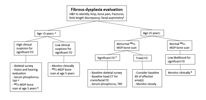
Suggested evaluations to determine if fibrous dysplasia (FD) is present and the extent of disease if FD is present

99Tc-MDP = technetium-99 conjugated with methylene diphosphonate; FD = fibrous dysplasia; H&P = history and physical exam; TRP = tubular reabsorption of phosphate; XR = x-ray

1. Performed at initial presentation in all individuals suspected of having FD/MAS.

2. Areas of clinically significant FD will be apparent on bone scan by age five years. Prior to age five years, a normal 99Tc-MDP does not eliminate the possibility of significant FD [Hart et al 2007].

3. FGF23-mediated phosphate wasting is associated with a high FD burden. Phosphate wasting may worsen during rapid skeletal growth and improve or resolve in adulthood as FD becomes less active [Riminucci et al 2003].

4. Consider performing 99Tc-MDP bone scan in children younger than age five years regardless of clinical suspicion for bone disease in instances where establishing the diagnosis of MAS may alter management – i.e., those for whom diagnostic surgery is being considered, such as skeletal biopsy.

5. Significance of FD is determined by both the amount and location of affected bone [Collins et al 2005]. Clinically significant disease is frequently associated with the craniofacial area, proximal femurs, and spine.

6. A normal 99Tc-MDP bone scan at age five years or older effectively eliminates clinically significant FD, and no further radiologic monitoring is required [Hart et al 2007].

From: Fibrous Dysplasia / McCune-Albright Syndrome

Copyright © 1993-2024, University of Washington, Seattle. GeneReviews is a registered trademark of the University of Washington, Seattle. All rights reserved.

GeneReviews® chapters are owned by the University of Washington. Permission is hereby granted to reproduce, distribute, and translate copies of content materials for noncommercial research purposes only, provided that (i) credit for source (http://www.genereviews.org/) and copyright (© 1993-2024 University of Washington) are included with each copy; (ii) a link to the original material is provided whenever the material is published elsewhere on the Web; and (iii) reproducers, distributors, and/or translators comply with the GeneReviews® Copyright Notice and Usage Disclaimer. No further modifications are allowed. For clarity, excerpts of GeneReviews chapters for use in lab reports and clinic notes are a permitted use.

For more information, see the GeneReviews® Copyright Notice and Usage Disclaimer.

For questions regarding permissions or whether a specified use is allowed, contact: ude.wu@tssamda.

Views

  • Cite this Page
  • PDF version of this page (1.3M)

Related information

  • OMIM
    Related OMIM records
  • PMC
    PubMed Central citations
  • PubMed
    Links to PubMed
  • Gene
    Locus Links

Similar articles in PubMed

See reviews...See all...

Recent Activity

Your browsing activity is empty.

Activity recording is turned off.

Turn recording back on

See more...