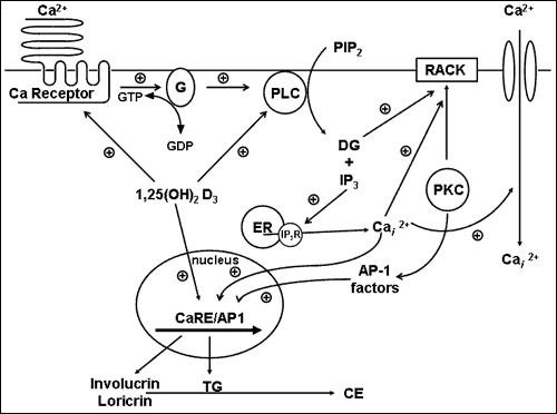
Figure 8. . A model of 1,25(OH)2D and calcium regulated keratinocyte differentiation.

Figure 8.

A model of 1,25(OH)2D and calcium regulated keratinocyte differentiation. The G-protein coupled calcium receptor (CaSR) when activated by extracellular calcium activates Gα as described in the legend to figure 6. Gα stimulates PLC mediated hydrolysis of PIP2 to IP3 and DG. IP3 releases Cai from intracellular stores, and DG activates PKC. Depletion of intracellular calcium stores leads to influx of calcium across store operated calcium channels. PKC stimulation leads to activation of AP-1 transcription factors which along with calcium and 1,25(OH)2D activated transcription factors stimulate the expression of genes essential for the differentiation process. 1,25(OH)2D regulates this process by inducing CaSR and PLC as well as genes essential for cornified envelope formation such as involucrin and transglutaminase.

From: Vitamin D: Production, Metabolism and Mechanisms of Action

Cover of Endotext
Endotext [Internet].
Feingold KR, Anawalt B, Blackman MR, et al., editors.
South Dartmouth (MA): MDText.com, Inc.; 2000-.
Copyright © 2000-2024, MDText.com, Inc.

This electronic version has been made freely available under a Creative Commons (CC-BY-NC-ND) license. A copy of the license can be viewed at http://creativecommons.org/licenses/by-nc-nd/2.0/.

NCBI Bookshelf. A service of the National Library of Medicine, National Institutes of Health.