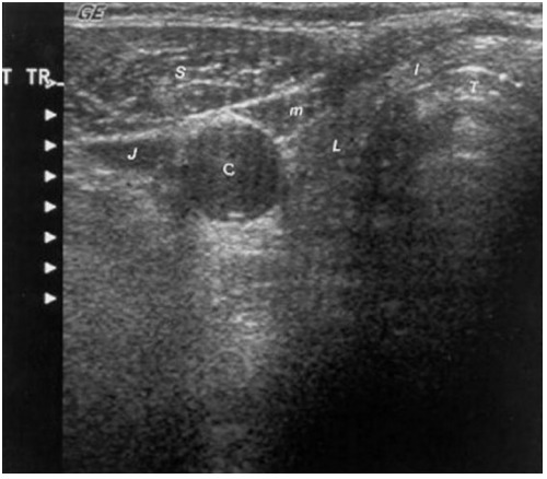
Figure 1. . Sonogram of the neck in the transverse plane showing a normal right thyroid lobe and isthmus.

Figure 1.

Sonogram of the neck in the transverse plane showing a normal right thyroid lobe and isthmus. L = small thyroid lobe in a patient who is taking suppressive amounts of L-thyroxine, I = isthmus, T = tracheal ring (the dense white arc represents calcification, distal to it reflects artifact), C = carotid artery (note the enhanced echoes deep to the fluid-filled blood vessel), J = jugular vein, S = sternocleidomastoid muscle, m = strap muscle.

From: Ultrasonography of the Thyroid

Cover of Endotext
Endotext [Internet].
Feingold KR, Anawalt B, Blackman MR, et al., editors.
South Dartmouth (MA): MDText.com, Inc.; 2000-.
Copyright © 2000-2024, MDText.com, Inc.

This electronic version has been made freely available under a Creative Commons (CC-BY-NC-ND) license. A copy of the license can be viewed at http://creativecommons.org/licenses/by-nc-nd/2.0/.

NCBI Bookshelf. A service of the National Library of Medicine, National Institutes of Health.