From: Chapter 30, Structural Biology of Glycan Recognition

PDF files are not available for download.
NCBI Bookshelf. A service of the National Library of Medicine, National Institutes of Health.

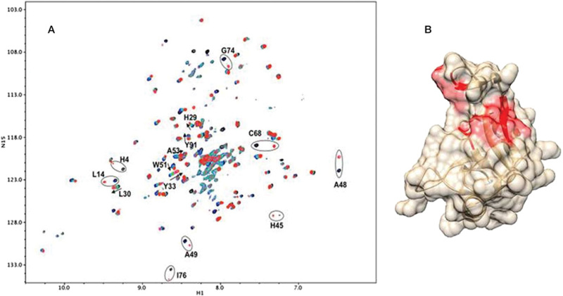
Chemical shift mapping of slow and fast exchange binding sites for a 4-sulfated chondroitin sulfate (CS) hexamer on the Link module of TSG6. (A) Cross peaks from spectra with increasing amounts of hexamer are superimposed. Those from residues experiencing fast exchange show progressive shifts and are marked with arrows; those from residues experiencing slow exchange show a pair of peaks, one appearing while another disappears, and are enclosed in ellipses. (B) Residues showing slow exchange are mapped in red on a crystal structure (2PF5).
Download Teaching Slide (PPTX, 1.8M)
From: Chapter 30, Structural Biology of Glycan Recognition

PDF files are not available for download.
NCBI Bookshelf. A service of the National Library of Medicine, National Institutes of Health.