Figure 2.

Figure 2.

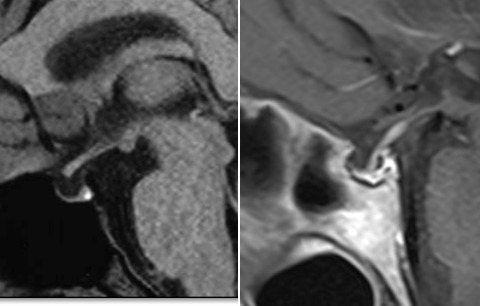
Sagittal T1W images of two patients with empty sella and Sheehan’s syndrome

Left Panel (sagittal T1W image without contrast) and Right (sagittal T1W image after contrast enhancement) represent the MRI appearance of empty sella in two patients with Sheehan’s syndrome. Both patients presented with hyponatremic coma due to unrecognized panhypopituitarism and infection 3 and 20 years after delivery complicated by postpartum hemorrhage.

From: Empty Sella

Cover of Endotext
Endotext [Internet].
Feingold KR, Anawalt B, Blackman MR, et al., editors.
South Dartmouth (MA): MDText.com, Inc.; 2000-.
Copyright © 2000-2024, MDText.com, Inc.

This electronic version has been made freely available under a Creative Commons (CC-BY-NC-ND) license. A copy of the license can be viewed at http://creativecommons.org/licenses/by-nc-nd/2.0/.

NCBI Bookshelf. A service of the National Library of Medicine, National Institutes of Health.