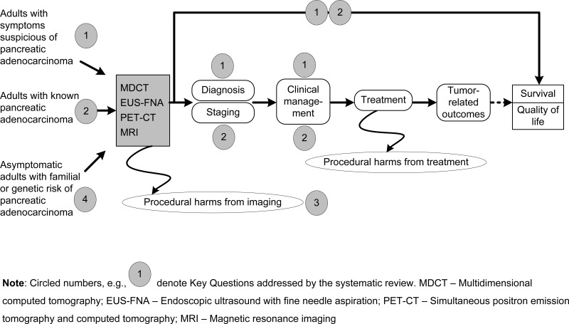
Figure 1 is an analytic framework intended to show how the four Key Questions fit within the general scope of imaging for pancreatic adenocarcinoma. The three relevant patient populations are listed in the left-hand column, and include adults with symptoms suspicious of pancreatic adenocarcinoma (Key Question 1), adults with known pancreatic adenocarcinoma (Key Question 3), and asymptomatic adults with familial or genetic risk of pancreatic adenocarcinoma (Key Question 4). These patients received one or more of the imaging modalities listed next (MDCT, MDCT with angiography, EUS-FNA, PET-CT, and MRI). AFter imaging tests, the figure proceeds to the right through diagnosis, staging, choices for clinical management, treatment (which may involve surgical resection if deemed appropriate), tumor-related outcomes, and patient-oriented outcomes including survival and quality of life. Key Question 3 involves the procedural harms from imaging, such as radiation dose from MDCT.

Figure 1Analytic framework

This figure depicts the Key Questions within the context of the PICOTS below. In general, the figure illustrates how different types of patients (the three populations listed on the left) can undergo different imaging tests (large box), resulting in the intermediate outcomes of diagnostic accuracy, staging accuracy, and clinical management decisions. Treatment is intended to improve (if possible) the patient-oriented outcomes listed to the right: survival and quality of life. Also, procedural harms of the imaging procedures may occur.

From: Introduction

Cover of Imaging Tests for the Diagnosis and Staging of Pancreatic Adenocarcinoma
Imaging Tests for the Diagnosis and Staging of Pancreatic Adenocarcinoma [Internet].
Comparative Effectiveness Review, No. 141.
Treadwell J, Mitchell M, Eatmon K, et al.

NCBI Bookshelf. A service of the National Library of Medicine, National Institutes of Health.