NCBI Bookshelf. A service of the National Library of Medicine, National Institutes of Health.

Adam MP, Feldman J, Mirzaa GM, et al., editors. GeneReviews® [Internet]. Seattle (WA): University of Washington, Seattle; 1993-2024.

Kagami-Ogata Syndrome

Synonym: KOS14
, MD, PhD
Department of Pediatrics
Hamamatsu University School of Medicine
Hamamatsu, Japan
, MD, PhD
Department of Molecular Endocrinology
National Research Institute for Child Health and Development
Tokyo, Japan

Initial Posting: .

Estimated reading time: 29 minutes

Summary

Clinical characteristics.

Kagami-Ogata syndrome is characterized by developmental delay, intellectual disability, feeding difficulty with impaired swallowing, full cheeks, prominent and deep philtrum, small bell-shaped thorax with coat-hanger appearance of the ribs, and abdominal wall defects (omphalocele and diastasis recti). Additional common features include joint contractures, kyphoscoliosis, coxa valga, and laryngomalacia. Cardiac disease and hepatoblastoma have also been reported.

Diagnosis/testing.

The diagnosis of Kagami-Ogata syndrome is established in a proband with suggestive findings and findings on molecular genetic testing that suggest deficient expression of the RTL1 antisense (RTL1as) allele of maternal origin due to one of the following: paternal uniparental disomy of chromosome 14; epimutation (hypermethylation) affecting the normally unmethylated MEG3/DLK1 intergenic differentially methylated region (MEG3/DLK1:IG-DMR) and MEG3 transcription start site DMR (MEG3:TSS-DMR) on the maternal allele; deletion of the maternally inherited 14q32.2 region that includes MEG3/DLK1:IG-DMR and/or MEG3:TSS-DMR, and may include RTL1as; deletion of the maternally inherited RTL1as but not MEG3/DLK1:IG-DMR or MEG3:TSS-DMR; translocation (or inversion) disrupting the integrity between the maternally inherited MEG3 promoter at the MEG3:TSS-DMR and RTL1as.

Management.

Treatment of manifestations: Developmental and educational support; mechanical ventilation and oxygen therapy as needed after birth; tracheostomy as required; monitor and treat upper and lower respiratory tract infections; treatment of scoliosis per orthopedist; treatment of joint contractures per rehabilitation medicine specialist; tube feeding typically required for poor weight gain; gastrostomy tube feeding as needed; feeding training; treatment of cardiac disease per cardiologist; standard treatment of hepatoblastoma with surgery and chemotherapy; social work and family support.

Surveillance: Monitor developmental progress, educational needs, for evidence of aspiration or respiratory insufficiency, progression of kyphoscoliosis and joint contractures, nutritional status, safety of oral intake, constipation, and family and care coordination needs at each visit. Echocardiogram annually; abdominal ultrasound and serum alpha-fetoprotein every three months until age three to four years.

Agents/circumstances to avoid: Avoid exposure to respiratory infections; individuals with Kagami-Ogata syndrome may develop respiratory failure with respiratory infections, especially during infancy.

Genetic counseling.

The recurrence risk of Kagami-Ogata syndrome is dependent on the genetic mechanism underlying deficient expression of the maternal RTL1as allele in the proband. In most affected individuals, the underlying genetic mechanism occurs as a de novo event and the recurrence risk to sibs is not increased. Less commonly, a parent of a proband has a predisposing genetic alteration associated with an increased recurrence risk to sibs. Recurrence of Kagami-Ogata syndrome has been reported in sibs with maternally derived deletions. If a deletion or translocation involving the chromosome 14q32.2 imprinted region has been identified in the proband, prenatal testing and preimplantation genetic testing are possible. Methylation testing of fetal DNA to examine abnormal methylation patterns of the DMRs (MEG3/DLK1:IG-DMR and MEG3:TSS-DMR) is not recommended; while DNA extracted from amniotic fluid is currently believed to provide the most reliable tissue source for evaluating fetal methylation status, false negative findings have been reported.

Diagnosis

No consensus clinical diagnostic criteria for Kagami-Ogata syndrome have been published.

Suggestive Findings

Kagami-Ogata syndrome should be suspected in individuals with the following clinical findings, especially specific (pathognomonic) findings in addition to characteristic but not specific findings and nonspecific findings [Kagami et al 2015, Ogata & Kagami 2016].

Specific (pathognomonic) findings

Characteristic but not specific findings

  • Abdominal wall defects such as omphalocele and diastasis recti
  • Placentomegaly
  • Polyhydramnios

Nonspecific findings

  • Craniofaciocervical features such as frontal bossing, hirsute forehead, blepharophimosis, depressed nasal bridge, anteverted nares, puckered lips, micrognathia, and short, webbed neck
  • Relatively large birth weight
  • Developmental delay (moderate to severe)
  • Feeding difficulty with impaired swallowing
  • Joint contractures
  • Kyphoscoliosis

Note: Characteristic face, small bell-shaped thorax with coat-hanger appearance of the ribs, and omphalocele can be observed on fetal ultrasound and MRI from the second trimester [Chen et al 2019, Igreja da Silva et al 2019, Molinet Coll et al 2021, Kuriki et al 2022].

Establishing the Diagnosis

The diagnosis of Kagami-Ogata syndrome is established in a proband with suggestive findings and findings on molecular genetic testing that suggest deficient expression of the RTL1 antisense (RTL1as) allele of maternal origin due to one of the following (see Table 1):

  • Paternal uniparental disomy of chromosome 14 (upd(14)pat) (~50% of affected individuals)
  • Epimutation (hypermethylation) affecting the normally unmethylated MEG3/DLK1 intergenic differentially methylated region (MEG3/DLK1:IG-DMR) and MEG3 transcription start site DMR (MEG3:TSS-DMR) of maternal origin (~25% of affected individuals)
  • Deletion of the maternally inherited 14q32.2 region that includes MEG3/DLK1:IG-DMR and/or MEG3:TSS-DMR, and may include RTL1as (~20% of affected individuals)
  • Deletion of the maternally inherited RTL1as but not MEG3/DLK1:IG-DMR or MEG3:TSS-DMR (~5% of affected individuals)
  • Translocation (or inversion) disrupting the integrity between the maternally inherited MEG3 promoter at the MEG3:TSS-DMR and RTL1as (rare)

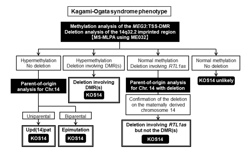
Molecular genetic testing for Kagami-Ogata syndrome includes recommended first-tier testing (methylation-specific multiple ligation-dependent probe amplification [MS-MLPA]) and second-tier testing (parent-of-origin analysis for chromosome 14) as needed to identify the molecular cause and clarify recurrence risk (see Figure 3).

First-Tier Testing

MS-MLPA that includes simultaneous methylation analysis of MEG3:TSS-DMR and deletion analysis of the multiple loci at the 14q32.2 imprinted region including MEG3/DLK1:IG-DMR, MEG3:TSS-DMR, and RTL1as is recommended. Note: (1) This testing can distinguish abnormal methylation caused by a deletion from abnormal methylation caused by upd(14)pat or epimutation, information that is important for recurrence risk counseling; (2) This testing can identify deletions of MEG3/DLK1:IG-DMR but not methylation abnormalities of MEG3/DLK1:IG-DMR, because the MS-MLPA does not address this methylation pattern.

  • If methylation is abnormal (identification of hypermethylation of MEG3:TSS-DMR) but no deletion is identified, second-tier testing (parent-of-origin testing for chromosome 14) can distinguish upd(14)pat from an epimutation.
  • If methylation is normal and a deletion of RTL1as is identified, second-tier testing can identify the parent of origin of the RTL1as deletion.

Second-Tier Testing

Parent-of-origin analysis for chromosome 14 using polymorphic DNA markers, which requires a DNA sample from the affected individual and both parents, can distinguish upd(14)pat and biparental chromosome 14 suggesting an epimutation. Note: Karyotype is recommended in individuals with upd(14)pat to identify those with robertsonian translocation or isochromosome 14q and clarify recurrence risk [Ogata & Kagami 2016].

Parent-of-origin testing for RTL1as deletion. Only a deletion of the maternal RTL1as allele results in Kagami-Ogata syndrome. Kagami-Ogata syndrome can be due to an RTL1as deletion inherited from the mother or a de novo deletion on chromosome 14 of maternal origin.

Other testing options (not recommended)

  • Methylation analysis (methylation-specific PCR, bisulfite sequencing, and pyrosequencing) can detect abnormal methylation pattern at 14q32.2 but cannot distinguish deletion from upd(14)pat and epimutation.
  • High-density SNP array analysis can detect smaller deletions and uniparental isodisomy of 14q32.2 but cannot detect uniparental heterodisomy of 14q32.2.

Table 1.

Molecular Genetic Testing Used in Kagami-Ogata Syndrome

MethodGenetic Mechanism 1Proportion of Probands Identified by Method 2
MS-MLPA 3upd(14)pat~50%
Epimutation (hypermethylation) of MEG3:TSS-DMR (maternal)~25%
Deletion of MEG3/DLK1:IG-DMR &/or MEG3:TSS-DMR (maternal)~20%
Deletion of RTL1as (maternal) 4~5% 5
KaryotypeTranslocations &/or inversions disrupting the integrity between the MEG3 promoter & RTL1asExtremely rare 6
Robertsonian translocations incl 14qRare

DMR = differentially methylated region; IG = intergenic; MS-MLPA = methylation-specific multiple ligation-dependent probe amplification; TSS = transcription start site; upd(14)pat = paternal uniparental disomy of chromosome 14

1.

See Molecular Genetics for more details and see Figure 4.

2.
3.

Identification of hypermethylation of MEG3:TSS-DMR establishes the diagnosis of Kagami-Ogata syndrome. However, deletion analysis (included in MS-MLPA) and parent-of-origin analysis are required to distinguish upd(14)pat, epimutation, and deletions involving the DMRs.

4.

Parent-of-origin analysis is necessary to confirm that the RTL1as deletion is present on the maternally inherited chromosome 14.

5.

Deletion of RTL1as (maternal) without deletion of MEG3/DLK1:IG-DMR or MEG3:TSS-DMR has been identified in five individuals with Kagami-Ogata syndrome. The frequency of such deletions may be overestimated, because of the publication bias that such a rare condition would be reported preferentially.

6.

A translocation disrupting the integrity between the maternally inherited MEG3 promoter at the MEG3:TSS-DMR and RTL1as has been identified in one individual with Kagami-Ogata syndrome.

Clinical Characteristics

Clinical Description

Kagami-Ogata syndrome is characterized by developmental delay, intellectual disability, feeding difficulty, full cheeks and prominent and deep philtrum, small bell-shaped thorax with coat-hanger appearance of the ribs, and abdominal wall defects (omphalocele and diastasis recti). Additional common features include joint contractures, kyphoscoliosis, coxa valga, and laryngomalacia. Cardiac disease and hepatoblastoma have also been reported [Kagami et al 2015, Ogata & Kagami 2016]. To date, approximately 100 individuals have been diagnosed with Kagami-Ogata syndrome [Sakaria et al 2021; Mackay et al 2022; Smith et al 2024; T Ogata & M Kagami, unpublished observations].

Table 2.

Kagami-Ogata Syndrome: Frequency of Select Features

Feature% of Persons w/Feature 1Comment
Pregnancy & delivery Polyhydramnios>95%
Placentomegaly~85%Placenta size >120% of normal
Development & cognition Developmental delay>95%Moderate to severe
Intellectual disability100%
Nutrition & growth Feeding difficulty>95%
Prenatal overgrowth>50%Birth length &/or weight >2 SD above mean
Postnatal growth deficiency~35%Height &/or weight >2 SD below mean
Craniofacial features Full cheeks & prominent & deep philtrum (most common & specific features)>90%-95%Additional features incl frontal bossing, hirsute forehead, blepharophimosis, depressed nasal bridge, anteverted nares, puckered (somewhat narrow & tented) lips, micrognathia, & short, webbed neck
Skeletal abnormalities Small bell-shaped thorax100%
Coat-hanger appearance of ribs100%
Joint contractures60%-65%
Kyphoscoliosis~40%
Coxa valga~33%
Respiratory Laryngomalacia~40%
Abdominal wall defects Diastasis recti65%-70%
Omphalocele~30%
Other features Cardiac disease25%
Hepatoblastoma5%-10%

SD = standard deviations

1.

Kagami et al [2015]; similar data have been summarized by Sakaria et al [2021].

Pregnancy and delivery. Polyhydramnios is typically identified in the second trimester at a median gestational age of 25.5 weeks (range: 14-30 weeks' gestation); amnioreduction is required in most pregnancies after 25 weeks' gestation (~80%) and in almost all pregnancies after 30 weeks' gestation. Polyhydramnios is due to placentomegaly and impaired swallowing. Thoracic and abdominal abnormalities are found on prenatal ultrasound in ~40% of fetuses from approximately 25 weeks' gestation. Premature delivery is frequent (~80%), with a median gestational age at delivery of 32.5 weeks (range: 30-35 weeks' gestation).

Developmental delay. Head control occurs at a median age of seven months (range: 3-36 months). Sitting without support occurs at a median age of 12 months (range: 8-25 months). Walking without support occurs at a median age of 25.5 months (range: 20-49 months). Only mild delay has been reported in two individuals with Kagami-Ogata syndrome: one individual with an epimutation [Higashiyama et al 2022] and one with mosaic paternal uniparental disomy of chromosome 14 (upd(14)pat) [Haug et al 2017].

Intellectual disability. Intellectual disability is reported in all individuals, with a median IQ of 55 (range: 29-70).

No abnormal brain MRI findings were delineated in five individuals examined.

Growth. Prenatal growth is well preserved and birth weight and length are usually above the mean. Range in birth weight is 0.1 standard deviations (SD) below the mean to 8.8 SD above the mean, and range in birth length is 1.7 SD below the mean to 3.0 SD above the mean. However, postnatal growth is frequently compromised primarily due to poor nutrition related to respiratory failure and feeding difficulty. In individuals age one to 15 years, range in weight is 6.0 SD below the mean to 4.0 SD above the mean, and range in length/height is 8.7 SD below the mean to 1.1 SD above the mean.

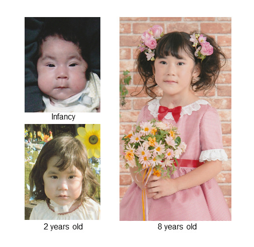
Craniofacial features (see Figure 1) in individuals with Kagami-Ogata syndrome include frontal bossing (~75%), hirsute forehead (~70%), blepharophimosis (70%-75%), full cheeks (>90%), depressed nasal bridge (>90%), anteverted nares (80%), prominent and deep philtrum (>95%), puckered (somewhat narrow and tented) lips (>50%), micrognathia (~100%), and short, webbed neck (>90%). Craniofacial features are typically present in infancy and consistently recognizable during childhood. While many of the craniofacial features are nonspecific, full cheeks and prominent and deep philtrum are very common and appear specific to Kagami-Ogata syndrome.

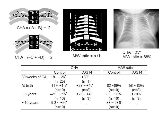
Thoracic abnormalities with respiratory failure. A small bell-shaped thorax is evident in infancy. Objective assessment of increased coat-hanger angle and decreased mid-to-widest thorax ratio is possible from fetal life through childhood (see Figure 2). The shape of the thorax appears normal after infancy, whereas coat-hanger appearance of the ribs remains discernible throughout childhood. Because of the thoracic anomalies, mechanical ventilation with oxygen has been required in more than 90% of individuals, with a median duration of one month (range: 0.1-17 months). Tracheostomy is sometimes necessary due to severe laryngomalacia.

Other skeletal abnormalities. Contractures at various joints, kyphoscoliosis, and coxa valga are also reported. The severity of these findings is variable among affected individuals.

Feeding/gastrointestinal manifestations. Tube feeding was required in all individuals who survived more than one week, with a median duration of 7.5 months (range: 0.1-89 months). Diastasis recti is common, and omphalocele can also occur. Abdominal wall weakness may cause constipation.

Cardiac disease. Cardiovascular malformations are recognized in ~25% of affected individuals. Of eight individuals who have been confirmed to have cardiac malformations, two have atrial septal defect, one has ventricular septal defect, four have patent ductus arteriosus, and one has pulmonary stenosis. These cardiac lesions are mild and do not require intensive medication or surgery. No individuals have been reported to have cardiomyopathy or conduction defects.

Hepatoblastoma has been identified in three individuals during infancy out of the 100 reported individuals to date.

Other. Simple seizure was reported in one individual [Kagami et al 2015].

Prognosis. Mortality is relatively high before age five years (20%-25% of individuals), but increased mortality is not observed in those with Kagami-Ogata syndrome older than age five years. The cause of death is variable; respiratory failure constitutes the primary cause of death. To date, nine adults age 18 years and older with Kagami-Ogata syndrome have been identified, including four unpublished individuals; the oldest affected individual is age 35 years [Smith et al 2024; T Ogata & M Kagami, personal observations].
Genotype-Phenotype Correlations

No clear genotype-phenotype correlations have been identified [Kagami et al 2015].

Note: Paternal uniparental disomy of chromosome 14, epimutations, and deletions that do not include RTL1as are associated with approximately five times the RTL1 expression compared to controls, whereas deletions including RTL1as are associated with approximately 2.5 times the RTL1 expression (see Molecular Genetics). However, because of the small number of deletions including RTL1as, it is unknown if the phenotype is milder in individuals with approximately 2.5 times the RTL1 expression than in individuals with five times the RTL1 expression compared to controls.

Penetrance

Penetrance is complete, and affected individuals invariably show full cheeks, prominent and deep philtrum, and small bell-shaped thorax with coat-hanger appearance of the ribs. However, there could be bias in that molecular studies are performed only when individuals have certain clinical features. In this regard, two individuals with upd(14)pat mosaicism have been reported to date; one individual has a mild phenotype, while the other has a typical Kagami-Ogata syndrome phenotype [Haug et al 2017, Li et al 2021]. Thus, some mosaic individuals with nearly normal phenotype may have been overlooked.

Nomenclature

Kagami-Ogata syndrome has also been referred to as upd(14)pat syndrome. However, the term upd(14)pat syndrome may be misleading, as Kagami-Ogata syndrome can be caused by genetic mechanisms other than paternal uniparental disomy of chromosome 14. Thus, "Kagami-Ogata syndrome" has been proposed to describe the unique clinical entity caused by various (epi)genetic aberrations affecting the chromosome 14q32.2 imprinted region.

Prevalence

To date, approximately 100 individuals with Kagami-Ogata syndrome have been reported [Mackay et al 2022, Eggermann et al 2023]. In Japan, 76 individuals with Kagami-Ogata syndrome (some of whom are reported in the literature) have been identified [T Ogata & M Kagami, personal observations]. It is likely that many individuals remain undiagnosed.
Differential Diagnosis

Table 3.

Disorders of Interest in the Differential Diagnosis of Kagami-Ogata Syndrome

Gene(s) / Genetic MechanismDisorderMOIFeatures of Disorder
Overlapping w/Kagami-Ogata syndromeDistinguishing from Kagami-Ogata syndrome
Abnormal methylation pattern at 11p15.5; CNV involving 11p15.5; or CDKN1C pathogenic variant Beckwith-Wiedemann syndrome See Footnote 1.
  • Polyhydramnios
  • Placentomegaly
  • Omphalocele
  • Overgrowth
  • Hepatoblastoma (↑ risk)
Absence of:
  • Characteristic face w/full cheeks & prominent & deep philtrum
  • Small bell-shaped thorax w/coat-hanger appearance of ribs
>20 genes incl:
DYNC2H1
IFT140
KIF7
NEK1
WDR19
WDR34
Short-rib thoracic dysplasia (formerly asphyxiating thoracic dysplasia—Jeune syndrome) (OMIM PS208500)ARSmall thorax
GPC3 Simpson-Golabi-Behmel syndrome type 1 XL
  • Polyhydramnios
  • Abdominal wall defects
  • Overgrowth
  • Hepatoblastoma (↑ risk)
Absence of small bell-shaped thorax w/coat-hanger appearance of ribs

AR = autosomal recessive; CNV = copy number variant; MOI = mode of inheritance; XL = X-linked

1.

Reliable recurrence risk assessment requires identification of the genetic mechanism in the proband that underlies the abnormal expression of imprinted genes in the BWS critical region. While the majority of families have a recurrence risk of less than 1%, certain underlying genetic mechanisms may be associated with a recurrence risk as high as 50% (see Beckwith-Wiedemann Syndrome).

Management

No clinical practice guidelines for Kagami-Ogata syndrome have been published. In the absence of published guidelines, the following recommendations are based on the authors' personal experience managing individuals with this disorder.

Evaluations Following Initial Diagnosis

To establish the extent of disease and needs in an individual diagnosed with Kagami-Ogata syndrome, the evaluations summarized in Table 4 (if not performed as part of the evaluation that led to the diagnosis) are recommended.

Table 4.

Kagami-Ogata Syndrome: Recommended Evaluations Following Initial Diagnosis

System/ConcernEvaluationComment
Development Developmental assessment
  • To incl gross motor & fine motor skills, adaptive, cognitive, & speech-language eval
  • Eval for early intervention / special education
Neurologic Neurologic eval
Respiratory Respiratory eval immediately after birthMost infants present w/respiratory failure due to upper or lower respiratory infection, esp during infancy & early childhood.
Musculoskeletal Orthopedics / physical medicine & rehab / PT & OT evalTo incl assessment of:
  • Contractures & kyphoscoliosis
  • Mobility, ADL, & need for adaptive devices
  • Need for PT (to improve gross motor skills) &/or OT (to improve fine motor skills)
Gastrointestinal/
Feeding
  • Gastroenterology / nutrition / feeding team eval
  • Assessment of height & weight
  • To incl eval of aspiration risk & nutritional status
  • Consider eval for gastrostomy tube placement in persons w/dysphagia &/or aspiration risk.
Cardiac disease Echocardiogram
Tumorigenesis Abdominal ultrasound for hepatoblastoma
Serum AFPSerum AFP level should be reviewed by a physician w/experience interpreting AFP levels if possible (e.g., oncologist, clinical geneticist)
Genetic counseling By genetics professionals 1To obtain a pedigree & inform affected persons & their families re nature, MOI, & implications of Kagami-Ogata syndrome to facilitate medical & personal decision making
Family support
& resources
By clinicians, wider care team, & family support organizationsAssessment of family & social structure to determine need for:

ADL = activities of daily living; AFP = alpha-fetoprotein; MOI = mode of inheritance; OT = occupational therapy; PT = physical therapy

1.

Medical geneticist, certified genetic counselor, certified advanced genetic nurse

Treatment of Manifestations

Supportive care to improve quality of life, maximize function, and reduce complications is recommended. This ideally involves multidisciplinary care by specialists in relevant fields (see Table 5).

Table 5.

Kagami-Ogata Syndrome: Treatment of Manifestations

Manifestation/ConcernTreatmentConsiderations/Other
Developmental delay /
Intellectual disability /
Neurobehavioral issues
See Developmental Delay / Intellectual Disability Management Issues.
Respiratory failure
  • Mechanical ventilation & oxygen therapy are almost invariably required immediately after birth.
  • Tracheostomy may be required.
  • Monitor for & treat upper & lower respiratory tract infections.
Skeletal abnormalities
  • Treatment of kyphoscoliosis per orthopedist; brace or surgery may be indicated.
  • Treatment per rehab medicine specialist for joint contractures
Poor weight gain /
Growth deficiency
  • Tube feeding is almost invariably required.
  • Gastrostomy tube feeding as needed
  • Feeding training/rehab is recommended.
Low threshold for clinical feeding eval &/or radiographic swallowing study when showing clinical signs or symptoms of dysphagia
Cardiac disease Treatment per cardiologistMedication may be indicated; there are no reports of persons w/Kagami-Ogata syndrome having cardiac surgery.
Hepatoblastoma Standard treatment w/surgical resection & chemotherapy
Family/Community Ensure appropriate social work involvement to connect families w/local resources, respite, & support.Ongoing assessment of need for palliative care involvement &/or home nursing

Developmental Delay / Intellectual Disability Management Issues

In the absence of knowledge about the detailed clinical course of Kagami-Ogata syndrome, careful follow up appropriate for each affected individual is recommended.

Motor Dysfunction

Gross motor dysfunction. Physical therapy is recommended to maximize mobility and reduce the risk for later-onset orthopedic complications (e.g., contractures, scoliosis, hip dislocation).

Fine motor dysfunction. Occupational therapy is recommended for difficulty with fine motor skills that affect adaptive function such as feeding, grooming, dressing, and writing.

Oral motor dysfunction should be assessed at each visit and clinical feeding evaluations and/or radiographic swallowing studies should be obtained for choking/gagging during feeds, poor weight gain, frequent respiratory illnesses, or feeding refusal that is not otherwise explained. Assuming that the child is safe to eat by mouth, feeding therapy (typically from an occupational or speech therapist) is recommended to help improve coordination or sensory-related feeding issues. Feeds can be thickened or chilled for safety. When feeding dysfunction is severe, a nasogastric or gastrostomy tube may be necessary.

Communication issues. Consider evaluation for alternative means of communication for individuals who have expressive language difficulties.

Neurobehavioral/Psychiatric Concerns

Consultation with a developmental pediatrician may be helpful in guiding parents through appropriate behavior management strategies.

Surveillance

To monitor existing manifestations, the individual's response to supportive care, and the emergence of new manifestations, the evaluations summarized in Table 6 are recommended.

Table 6.

Kagami-Ogata Syndrome: Recommended Surveillance

System/ConcernEvaluationFrequency
Development Monitor developmental progress & educational needs.At each visit
Respiratory Monitor for evidence of aspiration, respiratory insufficiency.
Skeletal abnormalities Monitor for progression of kyphoscoliosis & joint contractures.
Feeding Eval of nutritional status & safety of oral intake
Gastrointestinal Monitor for constipation.
Cardiac EchocardiogramAnnually
Hepatoblastoma
  • Abdominal ultrasound
  • Serum AFP
Every 3 mos until age 3-4 yrs 1, 2
Family/Community Assess family need for social work support (e.g., palliative/respite care, home nursing, other local resources), care coordination, or follow-up genetic counseling if new questions arise (e.g., family planning).At each visit

AFP = alpha-fetoprotein

1.
2.

This screening has been recommended for hepatoblastoma in Beckwith-Wiedemann syndrome [Kalish et al 2017, Kalish et al 2024].

Agents/Circumstances to Avoid

Avoid exposure to respiratory infections; individuals with Kagami-Ogata syndrome may develop respiratory failure with respiratory infections, especially during infancy.

Evaluation of Relatives at Risk

See Genetic Counseling for issues related to testing of at-risk relatives for genetic counseling purposes.

Therapies Under Investigation

Search ClinicalTrials.gov in the US and EU Clinical Trials Register in Europe for access to information on clinical studies for a wide range of diseases and conditions. Note: There may not be clinical trials for this disorder.

Genetic Counseling

Genetic counseling is the process of providing individuals and families with information on the nature, mode(s) of inheritance, and implications of genetic disorders to help them make informed medical and personal decisions. The following section deals with genetic risk assessment and the use of family history and genetic testing to clarify genetic status for family members; it is not meant to address all personal, cultural, or ethical issues that may arise or to substitute for consultation with a genetics professional. —ED.

Mode of Inheritance

The recurrence risk of Kagami-Ogata syndrome is dependent on the genetic mechanism underlying deficient expression of the maternal RTL1as allele in the proband. Genetic mechanisms known to be causative of Kagami-Ogata syndrome include the following:

  • Paternal uniparental disomy of chromosome 14 (upd(14)pat) *
  • Epimutation (hypermethylation) of the normally unmethylated MEG3/DLK1 intergenic differentially methylated region (MEG3/DLK1:IG-DMR) and MEG3 transcription start site DMR (MEG3:TSS-DMR) of maternal origin *
  • Deletion of the 14q32.2 region that includes MEG3/DLK1:IG-DMR and/or MEG3:TSS-DMR, and may include RTL1as *
  • Translocation (or inversion) disrupting the integrity between the maternally inherited MEG3 promoter at the MEG3:TSS-DMR and RTL1as

In most affected individuals, the underlying genetic mechanism occurs as a de novo event and the recurrence risk to sibs is not increased. Less commonly, a parent of the proband has a predisposing genetic alteration (e.g., a maternally transmitted deletion) associated with an increased recurrence risk to sibs.

* If the diagnosis of Kagami-Ogata syndrome in the proband has been established by identification of hypermethylation of MEG3/DLK1:IG-DMR and MEG3:TSS-DMR, deletion analysis (if not already performed in conjunction with methylation analysis via MS-MLPA) and parent-of-origin analysis are required to distinguish upd(14)pat, epimutation, and deletions involving the DMRs. Parent-of-origin analysis for chromosome 14 using polymorphic DNA markers (which requires a DNA sample from the proband and both parents) can distinguish upd(14)pat and biparental chromosome 14 suggesting an epimutation (see Establishing the Diagnosis and Figure 3).

Risk to Family Members

Parents of a proband

  • The parents of a proband with Kagami-Ogata syndrome are not affected with Kagami-Ogata syndrome but may have a predisposing genetic alteration associated with the disorder such the following:
    • Maternal deletion involving the DMRs (MEG3/DLK1:IG-DMR and/or MEG3:TSS-DMR) and, in some individuals, RTL1as. Recurrence of Kagami-Ogata syndrome has been reported in sibs with maternally derived deletions [Kagami et al 2008, Beygo et al 2015, Kagami et al 2015, Luk 2017].
      If a proband with Kagami-Ogata syndrome has a maternally inherited deletion, the mother with the same deletion is theoretically predicted to have Kagami-Ogata syndrome when the deletion is derived from her mother, or Temple syndrome when the deletion is derived from her father and involves DLK1 (deletion of DLK1 is a disease-causing factor for Temple syndrome but not for Kagami-Ogata syndrome) (see Genetically Related Disorders).
      Transmission of a deletion from a mother with Temple syndrome to a child with Kagami-Ogata syndrome has been reported in several families [Kagami et al 2008]. Transmission of a deletion from a mother with Kagami-Ogata syndrome to a child with Kagami-Ogata syndrome has not been reported to date.
    • Maternal translocation (or inversion) disrupting the integrity between the MEG3 promoter and RTL1as
    • Paternal or maternal robertsonian translocation involving chromosome 14 or isochromosome 14q (1(14q)).
  • Evaluation of the parents to clarify recurrence risk is recommended; specific testing recommendations depend on the genetic mechanism underlying Kagami-Ogata syndrome in the proband (see Sibs of a proband).

Sibs of a proband. The risk to sibs depends on the underlying genetic mechanism in the proband and the genetic status of the parents. If the genetic mechanism underlying Kagami-Ogata syndrome in the proband is:

  • Paternal uniparental disomy of chromosome 14. If the genetic mechanism underlying Kagami-Ogata syndrome in the proband is upd(14)pat, karyotype of the proband is recommended to identify those with a robertsonian translocation or i(14q) [Ogata & Kagami 2016]. If a robertsonian translocation or i(14q) is identified in the proband (or if the chromosomal status of the proband is unknown), parental karyotyping is recommended.
    • If both parents have normal karyotypes, it is presumed that upd(14)pat occurred in the proband as a de novo event and the recurrence risk to sibs is <1%.
    • If either of the parents has a robertsonian translocation or i(14q), the recurrence risk to sibs is increased. Note: While upd(14)pat mediated by parental robertsonian translocation has been identified in multiple individuals with Kagami-Ogata syndrome [Wang et al 2020], sib recurrence of upd(14)pat mediated by this mechanism has not been reported.
  • Epimutation (hypermethylation) of the maternally inherited DMRs. If the genetic mechanism underlying Kagami-Ogata syndrome in the proband is epimutation (hypermethylation) of the maternally inherited DMRs, it is presumed that hypermethylation occurred in the proband as a de novo event and the recurrence risk to sibs is <1%. Additional testing of the proband and the parents is not required to clarify recurrence risk. Note: (1) The underlying genetic mechanism of hypermethylation is unknown. (2) Kagami-Ogata syndrome associated with multilocus imprinting disturbance (MLID) has not been reported [Kagami et al 2017a].
  • Deletion involving the maternally inherited 14q32.2 imprinted region. If the genetic mechanism underlying Kagami-Ogata syndrome in the proband is deletion involving the maternally inherited 14q32.2 imprinted region, targeted deletion analysis is recommended for the mother of the proband.
    • If the deletion identified in the proband is not identified in maternal leukocyte DNA, it is presumed that the deletion occurred in the proband as a de novo event and the recurrence risk to sibs is <1%. Note: Testing of maternal leukocyte DNA may not detect all instances of somatic mosaicism and will not detect a deletion that is present in the germ (gonadal) cells only. (Gonadal mosaicism for a deletion involving the 14q32.2 imprinted region has not been reported to date.)
    • If the mother of the proband has the deletion, the risk to sibs is 50%. Recurrence of Kagami-Ogata syndrome has been reported in sibs with maternally derived deletions [Kagami et al 2008, Beygo et al 2015, Kagami et al 2015, Luk 2017]. (Note: If the deletion includes DLK1 and the mother inherited the deletion from her father, the mother likely has Temple syndrome.)
  • Translocation or inversion disrupting the integrity between the maternally inherited MEG3 promoter and RTL1as. If the genetic mechanism underlying Kagami-Ogata syndrome in the proband is translocation or inversion disrupting the integrity between the maternally inherited MEG3 promoter and RTL1as, karyotype analysis is recommended for the mother of the proband.
    • If the mother of the proband has a normal karyotype, it is presumed that the translocation (or inversion) occurred in the proband as a de novo event and the recurrence risk to sibs is <1%.
    • If the mother of the proband has the same translocation or inversion, the recurrence risk to sibs is increased.

Offspring of a proband. Many individuals with Kagami-Ogata syndrome are not yet of reproductive age and, to date, individuals with Kagami-Ogata syndrome are not known to reproduce. However, reproductive function is predicted to be preserved in Kagami-Ogata syndrome and the following recurrence risk issues should be considered:

  • If the proband has a deletion involving the 14q32.2 imprinted region, offspring of the proband are at risk for Kagami-Ogata syndrome or Temple syndrome depending on the sex of the proband and the deletion size. If the proband is a female, offspring are predicted to have a 50% risk of Kagami-Ogata syndrome. If the proband is male and the deletion encompasses DLK1, offspring are predicted to have a 50% risk of Temple syndrome.
  • If the proband has a chromosome alteration predicted to result in uniparental disomy in offspring (e.g., a robertsonian translocation or isochromosome 14q), offspring of the proband are at risk for upd(14)pat-mediated Kagami-Ogata syndrome or upd(14)mat-mediated Temple syndrome, depending on the sex of the proband and the underlying mechanism for the development of uniparental disomy (i.e., trisomy rescue or monosomy rescue).

Other family members

  • If the mother of the proband is heterozygous for a deletion involving the 14q32.2 imprinted region, the mother's sibs are at risk of having the deletion.
  • If a chromosome alteration involving 14q32.2 (e.g., a translocation) is identified in the mother of the proband, the mother's sibs are at risk of having the chromosome alteration.

Related Genetic Counseling Issues

Family planning

  • The optimal time for determination of genetic risk and discussion of the availability of prenatal/preimplantation genetic testing is before pregnancy.
  • It is appropriate to offer genetic counseling (including discussion of potential risks to offspring and reproductive options) to the parents of affected individuals.

Prenatal Testing and Preimplantation Genetic Testing

Positive Family History

Genomic variants

  • If a deletion or translocation involving the chromosome 14q32.2 imprinted region has been identified in the proband, prenatal testing using fetal DNA from samples obtained by chorionic villus sampling or amniocytes and preimplantation genetic testing are possible [Sasaki et al 2014, Sabria-Back et al 2022].
  • If the proband has Kagami-Ogata syndrome as the result of upd(14)pat (and chromosome analysis of both parents is normal) or an epimutation (hypermethylation), the recurrence risk is presumed to be <1% and prenatal/preimplantation genetic testing is likely unwarranted.

Methylation changes. Methylation testing of fetal DNA to examine abnormal methylation patterns of the DMRs (MEG3/DLK1:IG-DMR and MEG3:TSS-DMR) is not recommended. While DNA extracted from amniotic fluid is currently believed to provide the most reliable tissue source for evaluating fetal methylation status, false negative findings have been reported [Eggermann et al 2016]. Genetic counseling regarding the potential limitations of prenatal testing for epigenetic alterations should be undertaken [Eggermann et al 2016].

Prenatal findings

  • Even in the absence of obvious clinical findings of Kagami-Ogata syndrome on prenatal ultrasound investigation, a residual risk for recurrence of Kagami-Ogata syndrome remains given the variability in clinical presentation and the limitation of fetal ultrasound studies. A newborn at risk for Kagami-Ogata syndrome should be monitored for respiratory and feeding conditions.
  • Maternal serum alpha-fetoprotein concentration may be elevated from the second trimester in the presence of omphalocele [Campbell & Copel 2018].

Negative Family History

Prenatal testing may be considered when clinical features suggestive of Kagami-Ogata syndrome such as polyhydramnios, characteristic facies, small thorax with coat-hanger appearance of the ribs, and omphalocele are delineated in a fetus not known to be at risk for Kagami-Ogata syndrome. Prenatal testing performed in such conditions has revealed genetic abnormalities consistent with Kagami-Ogata syndrome, including mosaic upd(14)pat [Suzumori et al 2010, Chen et al 2019, Igreja da Silva et al 2019, Li et al 2021, Molinet Coll et al 2021, Kuriki et al 2022].

Differences in perspective may exist among medical professionals and within families regarding the use of prenatal and preimplantation genetic testing. While most health care professionals would consider use of prenatal and preimplantation genetic testing to be a personal decision, discussion of these issues may be helpful.

Resources

GeneReviews staff has selected the following disease-specific and/or umbrella support organizations and/or registries for the benefit of individuals with this disorder and their families. GeneReviews is not responsible for the information provided by other organizations. For information on selection criteria, click here.

No specific resources for Kagami-Ogata Syndrome have been identified by GeneReviews staff.

Molecular Genetics

Information in the Molecular Genetics and OMIM tables may differ from that elsewhere in the GeneReview: tables may contain more recent information. —ED.

Table B.

OMIM Entries for Kagami-Ogata Syndrome (View All in OMIM)

608149KAGAMI-OGATA SYNDROME

Molecular Pathogenesis

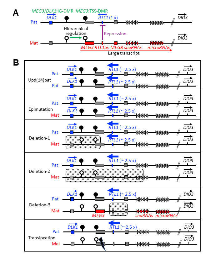
The primary underlying factor for the development of Kagami-Ogata syndrome is increased (~2.5 times or ~5 times that of controls) RTL1 expression due to absence of the functional RTL1 antisense (RTL1as) allele of maternal origin that acts as a trans-acting repressor for RTL1 expression [Kagami et al 2015, Ogata & Kagami 2016]. The maternal 14q32.2 region normally includes unmethylated MEG3/DLK1 intergenic differentially methylated region (MEG3/DLK1:IG-DMR) and unmethylated MEG3 transcription start site DMR (MEG3:TSS-DMR), resulting in normal expression of RTL1as. RTL1as expressed from the maternal allele represses RTL1 expression on the paternal allele (see Figure 4A).

Structure and character of the 14q32.2 imprinted region

  • This imprinted region contains the germline-derived MEG3/DLK1:IG-DMR and the postfertilization-derived MEG3:TSS-DMR; the protein-coding paternally expressed genes DLK1 and RTL1; and the non-coding maternally expressed genes MEG3, RTL1as, MEG8, and snoRNA and microRNA genes (see Figure 4A).
  • Both DMRs are methylated after paternal transmission and unmethylated after maternal transmission in the body; in the placenta, the MEG3/DLK1:IG-DMR remains as a DMR, but the MEG3:TSS-DMR is grossly hypomethylated regardless of parental origin.
  • The unmethylated MEG3:TSS-DMR and the MEG3/DLK1:IG-DMR of maternal origin function as imprinting control centers in the body and the placenta, respectively, and the MEG3:TSS-DMR can remain unmethylated only in the presence of unmethylated MEG3/DLK1:IG-DMR, because of the hierarchical regulation of the methylation patterns between the two DMRs [Kagami et al 2010].
  • All the non-coding maternally expressed genes are part of a large transcript expressed by the MEG3 promoter.

Mechanisms of disease causation include paternal uniparental disomy of chromosome 14 (upd(14)pat), epimutation (hypermethylation) of MEG3/DLK1:IG-DMR and MEG3:TSS-DMR of maternal origin, deletion of MEG3/DLK1:IG-DMR and/or MEG3:TSS-DMR of maternal origin, deletion of the maternally inherited RTL1as, or translocation (or inversion) disrupting the integrity between the maternally inherited MEG3 promoter at the MEG3:TSS-DMR and RTL1as.

Paternal uniparental disomy of chromosome 14. Advanced maternal age at childbirth is a risk factor for the development of monosomy rescue-mediated upd(14)pat [Kagami et al 2012].

Epimutation (hypermethylation) of the normally unmethylated MEG3/DLK1:IG-DMR and MEG3:TSS-DMR of maternal origin causes absence of functional RTL1as and results in a paternal methylation pattern.

  • Abnormal methylation of MEG3/DLK1:IG-DMR leads to abnormal methylation of MEG3:TSS-DMR on the maternally derived allele. There is no report describing hypermethylation of MEG3/DLK1:IG-DMR alone or MEG3:TSS-DMR alone.
  • The underlying cause of hypermethylation is unknown, and no multilocus imprinting disturbance (MLID) has been identified in individuals with Kagami-Ogata syndrome [Kagami et al 2017a].

Deletions including either DMRs

  • Deletions of MEG3/DLK1:IG-DMR and/or MEG3:TSS-DMR of maternal origin cause absence of functional RTL1as and a paternal methylation pattern [Kagami et al 2010].
  • Three different deletions involving maternally inherited MEG3:TSS-DMR alone have caused a paternal methylation pattern [Kagami et al 2010, Beygo et al 2015, Kilich et al 2024].
  • DMR deletions that include RTL1as result in ~2.5 times RTL1 expression compared to that of controls; DMR deletions that do not include RTL1as result in about five times RTL1 expression in the absence of functional RTL1as.
  • When deletions include DLK1, this leads to normal DLK1 expression, whereas when they do not include DLK1, this results in around two times the DLK1 expression seen in controls.
  • MEG genes are deleted or silenced.

Deletion of RTL1as (not including MEG3/DLK1:IG-DMR or MEG3:TSS-DMR)

  • This deletion results in loss of RTL1as expression from the maternally inherited chromosome 14q32.2 and resultant overexpression of RTL1 without altered methylation.
  • MEG8 is also deleted or disrupted in all deletions of this type reported to date.
  • DLK1 and non-deleted maternally expressed genes are predicted to be expressed normally.

Translocation disrupting the integrity between the MEG3 promoter and RTL1as silences transcription of RTL1as.

Chapter Notes

Author Notes

Tsutomu Ogata (pj.ca.dem-amah@atagomot) and Masayo Kagami (pj.og.dhccn@sm-imagak) are actively involved in clinical research regarding individuals with Kagami-Ogata syndrome. They would be happy to communicate with persons who have any questions regarding diagnosis of Kagami-Ogata syndrome or other considerations.

National Research Institute for Child Health and Development

European Network for Human Congenital Imprinting Disorders

Acknowledgments

This work was supported by the grant from Japan Agency for Medical Research and Development (AMED) (24ek0109587).

Revision History

  • 24 October 2024 (sw) Review posted live
  • 29 February 2024 (to) Original submission

References

Literature Cited

  • Beygo J, Elbracht M, de Groot K, Begemann M, Kanber D, Platzer K, Gillessen-Kaesbach G, Vierzig A, Green A, Heller R, Buiting K, Eggermann T. Novel deletions affecting the MEG3-DMR provide further evidence for a hierarchical regulation of imprinting in 14q32. Eur J Hum Genet. 2015;23:180-8. [PMC free article: PMC4297900] [PubMed: 24801763]
  • Campbell KH, Copel JA. Omphalocele. In: Copel JA, D’Alton ME, Feltovich H, Gratacos E, Krakow D, Odibo AO, Platt LD, Tuuschck B, eds. Obstetric Imaging: Fetal Diagnosis and Care. 2nd ed. Amsterdam: Elsevier; 2018:84–91.
  • Chen CL, Lee CN, Lin MW, Hsu WW, Tai YY, Lin SY. Prenatal diagnosis of paternal uniparental disomy for chromosome 14 using a single-nucleotide-polymorphism-based microarray analysis: a case report. J Formos Med Assoc. 2019;118:739-42. [PubMed: 30616993]
  • Eggermann T, Brioude F, Russo S, Lombardi MP, Bliek J, Maher ER, Larizza L, Prawitt D, Netchine I, Gonzales M, Grønskov K, Tümer Z, Monk D, Mannens M, Chrzanowska K, Walasek MK, Begemann M, Soellner L, Eggermann K, Tenorio J, Nevado J, Moore GE, Mackay DJ, Temple K, Gillessen-Kaesbach G, Ogata T, Weksberg R, Algar E, Lapunzina P. Prenatal molecular testing for Beckwith-Wiedemann and Silver-Russell syndromes: a challenge for molecular analysis and genetic counseling. Eur J Hum Genet. 2016;24:784-93. [PMC free article: PMC4867462] [PubMed: 26508573]
  • Eggermann T, Monk D, de Nanclares GP, Kagami M, Giabicani E, Riccio A, Tümer Z, Kalish JM, Tauber M, Duis J, Weksberg R, Maher ER, Begemann M, Elbracht M. Imprinting disorders. Nat Rev Dis Primers. 2023;9:33. [PubMed: 37386011]
  • Haug MG, Brendehaug A, Houge G, Kagami M, Ogata T. Mosaic upd(14)pat in a patient with mild features of Kagami-Ogata syndrome. Clin Case Rep. 2017;6:91-5. [PMC free article: PMC5771875] [PubMed: 29375845]
  • Higashiyama H, Ohsone Y, Takatani R, Futatani T, Kosaki R, Kagami M. Two infants with mild, atypical clinical features of Kagami-Ogata syndrome caused by epimutation. Eur J Med Genet. 2022;65:104580. [PubMed: 35953028]
  • Igreja da Silva JI, Ribeiro B, Cadilhe A, Nogueira-Silva C. Paternal uniparental disomy for chromosome 14: prenatal management. BMJ Case Rep. 2019;12:e231705. [PMC free article: PMC6954787] [PubMed: 31892621]
  • Ioannides Y, Lokulo-Sodipe K, Mackay DJ, Davies JH, Temple IK. Temple syndrome: improving the recognition of an underdiagnosed chromosome 14 imprinting disorder: an analysis of 51 published cases. J Med Genet. 2014;51:495-501. [PubMed: 24891339]
  • Kagami M, Kato F, Matsubara K, Sato T, Nishimura G, Ogata T. Relative frequency of underlying genetic causes for the development of UPD(14)pat-like phenotype. Eur J Hum Genet. 2012;20:928-32. [PMC free article: PMC3421115] [PubMed: 22353941]
  • Kagami M, Kurosawa K, Miyazaki O, Ishino F, Matsuoka K, Ogata T. Comprehensive clinical studies in 34 patients with molecularly defined UPD(14)pat and related conditions (Kagami-Ogata syndrome). Eur J Hum Genet. 2015;23:1488-98. [PMC free article: PMC4613461] [PubMed: 25689926]
  • Kagami M, Matsubara K, Nakabayashi K, Nakamura A, Sano S, Okamura K, Hata K, Fukami M, Ogata T. Genome-wide multilocus imprinting disturbance analysis in Temple syndrome and Kagami-Ogata syndrome. Genet Med. 2017a;19:476-82. [PMC free article: PMC5392596] [PubMed: 27632690]
  • Kagami M, Nagasaki K, Kosaki R, Horikawa R, Naiki Y, Saitoh S, Tajima T, Yorifuji T, Numakura C, Mizuno S, Nakamura A, Matsubara K, Fukami M, Ogata T. Temple syndrome: comprehensive molecular and clinical findings in 32 Japanese patients. Genet Med. 2017b;19:1356-66. [PMC free article: PMC5729347] [PubMed: 28640239]
  • Kagami M, O'Sullivan MJ, Green AJ, Watabe Y, Arisaka O, Masawa N, Matsuoka K, Fukami M, Matsubara K, Kato F, Ferguson-Smith AC, Ogata T. The IG-DMR and the MEG3-DMR at human chromosome 14q32.2: hierarchical interaction and distinct functional properties as imprinting control centers. PLoS Genet. 2010;6:e1000992. [PMC free article: PMC2887472] [PubMed: 20585555]
  • Kagami M, Sekita Y, Nishimura G, Irie M, Kato F, Okada M, Yamamori S, Kishimoto H, Nakayama M, Tanaka Y, Matsuoka K, Takahashi T, Noguchi M, Tanaka Y, Masumoto K, Utsunomiya T, Kouzan H, Komatsu Y, Ohashi H, Kurosawa K, Kosaki K, Ferguson-Smith AC, Ishino F, Ogata T. Deletions and epimutations affecting the human 14q32.2 imprinted region in individuals with paternal and maternal upd(14)-like phenotypes. Nat Genet. 2008;40:237-42. [PubMed: 18176563]
  • Kalish JM, Becktell KD, Bougeard G, Brodeur GM, Diller LR, Doria AS, Hansford JR, Klein SD, Kohlmann WK, Kratz CP, MacFarland SP, Pajtler KW, Rednam SP, Schienda J, States LJ, Villani A, Weksberg R, Zelley K, Tomlinson GE, Brzezinski JJ. Update on surveillance for Wilms tumor and hepatoblastoma in Beckwith-Wiedemann syndrome and other predisposition syndromes. Clin Cancer Res. 2024. Epub ahead of print. [PMC free article: PMC11611621] [PubMed: 39320341]
  • Kalish JM, Doros L, Helman LJ, Hennekam RC, Kuiper RP, Maas SM, Maher ER, Nichols KE, Plon SE, Porter CC, Rednam S, Schultz KAP, States LJ, Tomlinson GE, Zelley K, Druley TE. Surveillance recommendations for children with overgrowth syndromes and predisposition to Wilms tumors and hepatoblastoma. Clin Cancer Res. 2017;23:e115-e122. [PMC free article: PMC5538793] [PubMed: 28674120]
  • Kilich G, Hassey K, Behrens EM, Falk M, Vanderver A, Rader DJ, Cahill PJ, Raper A, Zhang Z, Westerfer D, Jadhav T, Conlin L, Izumi K, Rajagopalan R, Sullivan KE; UDN Consortium. Kagami Ogata syndrome: a small deletion refines critical region for imprinting. NPJ Genom Med. 2024;9:5. [PMC free article: PMC10784583] [PubMed: 38212313]
  • Kuriki A, Hosoya S, Ozawa K, Wada S, Kosugi Y, Wada YS, Sekizawa A, Miyazaki O, Kagami M, Sago H. Quantitative assessment of coat-hanger ribs detected on three-dimensional ultrasound for prenatal diagnosis of Kagami-Ogata syndrome. J Obstet Gynaecol Res. 2022;48:3314-8. [PMC free article: PMC10087373] [PubMed: 36087043]
  • Li F, Liu S, Jia B, Wu R, Chang Q. Prenatal diagnosis of a mosaic paternal uniparental disomy for chromosome 14: a case report of Kagami-Ogata syndrome. Front Pediatr. 2021;9:691761. [PMC free article: PMC8566877] [PubMed: 34746047]
  • Luk HM. Familial Kagami-Ogata syndrome in Chinese. Clin Dysmorphol. 2017;26:124-7. [PubMed: 27753651]
  • Mackay D, Bliek J, Kagami M, Tenorio-Castano J, Pereda A, Brioude F, Netchine I, Papingi D, de Franco E, Lever M, Sillibourne J, Lombardi P, Gaston V, Tauber M, Diene G, Bieth E, Fernandez L, Nevado J, Tümer Z, Riccio A, Maher ER, Beygo J, Tannorella P, Russo S, de Nanclares GP, Temple IK, Ogata T, Lapunzina P, Eggermann T. First step towards a consensus strategy for multi-locus diagnostic testing of imprinting disorders. Clin Epigenetics. 2022;14:143. [PMC free article: PMC9641836] [PubMed: 36345041]
  • Miyazaki O, Nishimura G, Kagami M, Ogata T. Radiological evaluation of dysmorphic thorax of paternal uniparental disomy 14. Pediatr Radiol. 2011;41:1013-9. [PubMed: 21607596]
  • Molinet Coll C, Sabrià Bach J, Izquierdo Renau M, Alarcón Allen A, Monk D, Gómez Del Rincón O, Milà Recasens M, Martínez Crespo JM. Prenatal diagnosis of Kagami-Ogata syndrome. J Clin Ultrasound. 2021;49:498-501. [PubMed: 33179779]
  • Ogata T, Kagami M. Kagami-Ogata syndrome: a clinically recognizable upd(14)pat and related disorder affecting the chromosome 14q32.2 imprinted region. J Hum Genet. 2016;61:87-94. [PMC free article: PMC4771937] [PubMed: 26377239]
  • Omark J, Masunaga Y, Hannibal M, Shaw B, Fukami M, Kato F, Saitsu H, Kagami M, Ogata T. Kagami-Ogata syndrome in a patient with 46,XX,t(2;14)(q11.2;q32.2)mat disrupting MEG3. J Hum Genet. 2021;66:439-43. [PubMed: 33067531]
  • Prasasya R, Grotheer KV, Siracusa LD, Bartolomei MS. Temple syndrome and Kagami-Ogata syndrome: clinical presentations, genotypes, models and mechanisms. Hum Mol Genet. 2020 Sep 30;29(R1):R107-R116. [PMC free article: PMC8325017] [PubMed: 32592473]
  • Sabria-Back J, Monteagudo-Sánchez A, Sánchez-Delgado M, Ferguson-Smith AC, Gómez O, Pertierra Cartada A, Tenorio J, Nevado J, Lapunzina P, Pereda Aguirre A, Giménez Sevilla C, Toro Toro E, Perez de Nanclares G, Monk D. Preimplantation genetic testing for a chr14q32 microdeletion in a family with Kagami-Ogata syndrome and Temple syndrome. J Med Genet. 2022;59:253-61. [PubMed: 33579810]
  • Sakaria RP, Mostafavi R, Miller S, Ward JC, Pivnick EK, Talati AJ. Kagami-Ogata syndrome: case series and review of literature. AJP Rep. 2021;11:e65-e75. [PMC free article: PMC8159623] [PubMed: 34055463]
  • Sasaki A, Sumie M, Wada S, Kosaki R, Kurosawa K, Fukami M, Sago H, Ogata T, Kagami M. Prenatal genetic testing for a microdeletion at chromosome 14q32.2 imprinted region leading to UPD(14)pat-like phenotype. Am J Med Genet A. 2014;164A:264-6. [PubMed: 24259232]
  • Smith CS, Riddell M, Badalato L, Au PYB. Adults with paternal UPD14 causing Kagami-Ogata syndrome: Case report and review of the literature. Am J Med Genet A. 2024;194:e63625. [PubMed: 38741340]
  • Suzumori N, Ogata T, Mizutani E, Hattori Y, Matsubara K, Kagami M, Sugiura-Ogasawara M. Prenatal findings of paternal uniparental disomy 14: delineation of further patient. Am J Med Genet A. 2010;152A:3189-92. [PubMed: 21108407]
  • Wang X, Pang H, Shah BA, Gu H, Zhang L, Wang H. A male case of Kagami-Ogata syndrome caused by paternal unipaternal disomy 14 as a result of a robertsonian translocation. Front Pediatr. 2020;8:88. [PMC free article: PMC7076151] [PubMed: 32211354]

Figures

Figure 1. . Female at infancy, age two years, and age eight years with Kagami-Ogata syndrome due to a maternally inherited microdeletion of DLK1, MEG3/DLK1:IG-DMR, MEG3:TSS-DMR, MEG3, RTL1/RTL1as, MEG8, and a centromeric part of the snoRNA genes.

Figure 1.

Female at infancy, age two years, and age eight years with Kagami-Ogata syndrome due to a maternally inherited microdeletion of DLK1, MEG3/DLK1:IG-DMR, MEG3:TSS-DMR, MEG3, RTL1/RTL1as, MEG8, and a centromeric part of the snoRNA genes. Characteristic full cheeks and prominent and deep philtrum are observed from infancy through childhood.

Adapted from Ogata & Kagami [2016]

Figure 2. . Chest radiograph of a Japanese neonate with Kagami-Ogata syndrome.

Figure 2.

Chest radiograph of a Japanese neonate with Kagami-Ogata syndrome. The coat-hanger angle (CHA) is increased from 30 weeks' gestation through age five years in individuals with Kagami-Ogata syndrome. CHA is the average of the right and left angles between the horizontal axis at the sixth costovertebral junction and the peak point of the sixth posterior rib. Note: If the ribs slope downward, the midpoint of the sixth rib is used for CHA measurement. The mid-to-widest thorax ratio is decreased in infants and young children with Kagami-Ogata syndrome.

CHA = coat-hanger angle; GA = gestational age; KOS14 = Kagami-Ogata syndrome; M/W ratio = mid-to-widest thorax ratio

Adapted from Kagami et al [2015] and Ogata & Kagami [2016]

Figure 3.

Figure 3.

Testing algorithm to establish the diagnosis and molecular cause of Kagami-Ogata syndrome

Chr. = chromosome; DMR = differentially methylated region; KOS14 = Kagami-Ogata syndrome; MS-MLPA = methylation-specific multiple ligation-dependent probe amplification; RTL1as = RTL1 antisense; TSS = transcription start site; upd(14)pat = paternal uniparental disomy of chromosome 14

Figure 4. . Chromosome 14q32.

Figure 4.

Chromosome 14q32.2 imprinted region and representative genetic causes leading to Kagami-Ogata syndrome. The black and white circles represent methylated and unmethylated differentially methylated regions (DMRs), respectively.

A. Structure and character of the imprinted region

B. Expression dosage of imprinted genes in several conditions. Stippled areas denote deleted region, and the cutting mark indicates the translocation breakpoint.

Copyright © 1993-2024, University of Washington, Seattle. GeneReviews is a registered trademark of the University of Washington, Seattle. All rights reserved.

GeneReviews® chapters are owned by the University of Washington. Permission is hereby granted to reproduce, distribute, and translate copies of content materials for noncommercial research purposes only, provided that (i) credit for source (http://www.genereviews.org/) and copyright (© 1993-2024 University of Washington) are included with each copy; (ii) a link to the original material is provided whenever the material is published elsewhere on the Web; and (iii) reproducers, distributors, and/or translators comply with the GeneReviews® Copyright Notice and Usage Disclaimer. No further modifications are allowed. For clarity, excerpts of GeneReviews chapters for use in lab reports and clinic notes are a permitted use.

For more information, see the GeneReviews® Copyright Notice and Usage Disclaimer.

For questions regarding permissions or whether a specified use is allowed, contact: ude.wu@tssamda.

Bookshelf ID: NBK608430PMID: 39446997