From: What Is Osteoporosis?

NCBI Bookshelf. A service of the National Library of Medicine, National Institutes of Health.

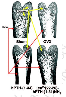
Typical specimens of demineralized (i.e., the calcium apatite was acid-extracted) distal femurs of OVXed (ovariectomized) rats showing that injecting 6.0 nmoles of PTH-(1-34)/1 kg of body weight once a day from the end of the 2nd week to the end of the 8th week after the operation did not stop the destruction and loss of trabeculae, while injecting the same molar dose of [Leu27]cyclo[Glu22-Lys26]hPTH-(1-31)NH2 (Ostabolin C™) reduced the loss of trabeculae. However, by 6 weeks of injections, both fragments had nearly doubled (1.8-2.0) the thickness of the remaining trabeculae compared to the mean thickness of the femurs in the trabeculae in the femurs of vehicle-injected OVXed control rats. The specimens were prepared at the end of the 6th week of the series of injections (i.e., 8 weeks after OVX). The lines—red for cortical bone and yellow for trabecular or cancellous—on these photographs are meant to acquaint the reader with the universal basic bone anatomy of a long bone such as the femur. The reader should also consult Kerr (1999) and Netter (1997) to learn the gross and microscopic anatomies of the various parts of the human skeleton. A color version of this figure is available online at http://www.Eurekah.com.
From: What Is Osteoporosis?

NCBI Bookshelf. A service of the National Library of Medicine, National Institutes of Health.