U.S. flag

An official website of the United States government

NCBI Bookshelf. A service of the National Library of Medicine, National Institutes of Health.

Adam MP, Feldman J, Mirzaa GM, et al., editors. GeneReviews® [Internet]. Seattle (WA): University of Washington, Seattle; 1993-2024.

Cover of GeneReviews®

GeneReviews® [Internet].

Show details

CYLD Cutaneous Syndrome

Synonyms: Brooke-Spiegler Syndrome (BSS), Familial Cylindromatosis (FC), Multiple Familial Trichoepithelioma (MFT)

, BSc, MBChB and , MBBS, PhD.

Author Information and Affiliations

Initial Posting: .

Estimated reading time: 26 minutes

Summary

Clinical characteristics.

CYLD cutaneous syndrome (CCS) typically manifests in the second or third decade with the appearance of multiple skin tumors including cylindromas, spiradenomas, trichoepitheliomas, and rarely, membranous basal cell adenoma of the salivary gland. The first tumor typically develops at puberty and tumors progressively accumulate through adulthood. Females often have more tumors than males. Tumors typically arise on the scalp and face but can also arise on the torso and sun-protected sites, such as the genital and axillary skin. A minority of individuals develop salivary gland tumors. Rarely, pulmonary cylindromas can develop in large airways and compromise breathing. Although the tumors are usually benign, malignant transformation is recognized.

Diagnosis / testing.

The diagnosis of CYLD cutaneous syndrome is established in a proband with multiple skin tumors (histologically confirmed cylindromas, spiradenomas, and/or trichoepitheliomas) and/or by identification of a germline heterozygous pathogenic variant in CYLD by molecular genetic testing.

Management.

Treatment of manifestations: Removal of cylindromas, spiradenomas, and trichoepitheliomas is by conventional surgery. Ideally, as much normal scalp and skin should be preserved. "Scalp-sparing" strategies include early primary excision with direct skin closure, tumor enucleation followed by direct skin closure, and excision followed by secondary intention healing techniques. Hyfrecation or laser ablation of selected small tumors may be considered. Mohs micrographic surgery for recurrence of tumors after failure of primary surgical excision may have limited benefit. Multidisciplinary team management of tumors that have undergone malignant transformation is recommended.

Prevention of primary manifestations: Appropriate precautions against UV-related skin damage are recommended.

Surveillance: Annual or more frequent full skin examination by a dermatologist, with assessment of tumor burden and rate of new tumor development, and for signs/symptoms of malignant transformation (rapid tumor growth, bleeding, ulceration, or appearance that is different from an affected individual's usual tumors).

Agents/circumstances to avoid: Radiotherapy should be avoided as it causes DNA damage and may result in further tumor formation or malignant transformation of existing lesions.

Evaluation of relatives at risk: It is appropriate to clarify the genetic status of apparently asymptomatic older and younger at-risk relatives of an affected individual in order to identify as early as possible those who would benefit from prompt initiation of treatment and preventive measures.

Genetic counseling.

Germline pathogenic variants in CYLD are inherited in an autosomal dominant manner. Most individuals with CYLD cutaneous syndrome inherit it from a parent. The degree of severity can vary within families; for example, a mildly affected parent may have a more severely affected child or vice versa. Offspring of an individual with CYLD cutaneous syndrome have a 50% chance of inheriting the variant. Prenatal testing for a pregnancy at increased risk is possible if the CYLD pathogenic variant in the family is known; however, requests for prenatal testing for later-onset diseases are uncommon and require careful genetic counseling.

Diagnosis

Formal diagnostic criteria for CYLD cutaneous syndrome (CCS) have not been established.

Suggestive Findings

CYLD cutaneous syndrome should be suspected in an individual with the following findings:

  • The presence of one or more cylindromas or spiradenomas on the face and scalp, perinasal trichoepitheliomas, or a combination of these tumor types in an individual
    Cylindromas, spiradenomas, and trichoepitheliomas can be diagnosed clinically but may mimic other skin tumors, thus requiring confirmatory skin biopsy.
  • A cylindroma or spiradenoma on the scalp or torso incidentally identified during an imaging study (CT, MRI, and/or PET scan) [Serra et al 1996, Brown et al 2018a]
  • A membranous basal cell adenoma-type salivary gland tumor in an individual with a single cylindroma, spiradenoma, or trichoepithelioma

Clinical genetic testing for a germline heterozygous CYLD pathogenic variant should be considered in an individual with the following [Dubois et al 2015]:

  • Two or more biopsy-confirmed cylindromas, spiradenomas, or trichoepitheliomas
  • A single biopsy-confirmed cylindroma, spiradenoma, or trichoepithelioma in the setting of a first-degree relative who has any one of these biopsy-confirmed tumors

Establishing the Diagnosis

The diagnosis of CYLD cutaneous syndrome is established in a proband with multiple skin tumors (histologically confirmed cylindromas, spiradenomas, and/or trichoepitheliomas) and/or by identification of a germline heterozygous pathogenic variant in CYLD by molecular genetic testing [Dubois et al 2015] (see Table 1).

When the phenotypic and laboratory findings suggest the diagnosis of CYLD cutaneous syndrome, molecular genetic testing approaches include single-gene testing or use of a multigene panel.

Single-gene testing. Sequence analysis of CYLD detects small intragenic deletions/insertions and missense, nonsense, and splice site variants; typically, exon or whole-gene deletions/duplications are not detected. Perform sequence analysis on a peripheral blood sample first.

A hereditary cancer multigene panel that includes CYLD and other genes of interest (see Differential Diagnosis) may also be considered. Note: (1) The genes included in the panel and the diagnostic sensitivity of the testing used for each gene vary by laboratory and are likely to change over time. (2) Some multigene panels may include genes not associated with the condition discussed in this GeneReview. (3) In some laboratories, panel options may include a custom laboratory-designed panel and/or custom phenotype-focused exome analysis that includes genes specified by the clinician. (4) Methods used in a panel may include sequence analysis, deletion/duplication analysis, and/or other non-sequencing-based tests.

For an introduction to multigene panels click here. More detailed information for clinicians ordering genetic tests can be found here.

Table 1.

Molecular Genetic Testing Used in CYLD Cutaneous Syndrome

Gene 1MethodProportion of Probands with a Pathogenic Variant 2 Detectable by Method
CYLD Sequence analysis 340%-100% 4, 5, 6
Gene-targeted deletion/duplication analysis 7Rare 8
1.
2.

See Molecular Genetics for information on allelic variants detected in this gene.

3.

Sequence analysis detects variants that are benign, likely benign, of uncertain significance, likely pathogenic, or pathogenic. Variants may include small intragenic deletions/insertions and missense, nonsense, and splice site variants; typically, exon or whole-gene deletions/duplications are not detected. For issues to consider in interpretation of sequence analysis results, click here.

4.

A single test center performing CYLD testing over a five-year period detected pathogenic CYLD variants in ∼70% of 56 probands who fulfilled previously published testing criteria using Sanger sequencing of coding exons [ESHG 2019].

5.

In a smaller study of 25 probands, the presence of cylindromas raised the likelihood of pathogenic variant detection to between 86% and 100% [Saggar et al 2008].

6.

In probands with only trichoepitheliomas, the rate of pathogenic variant detection was as low as 40% [Saggar et al 2008].

7.

Gene-targeted deletion/duplication analysis detects intragenic deletions or duplications. Methods used may include a range of techniques such as quantitative PCR, long-range PCR, multiplex ligation-dependent probe amplification (MLPA), and a gene-targeted microarray designed to detect single-exon deletions or duplications.

8.

Vanecek et al [2014] reported two affected individuals with large deletions of CYLD out of 13 affected individuals who did not demonstrate a pathogenic CYLD variant using Sanger sequencing.

Clinical Characteristics

Clinical Description

CYLD cutaneous syndrome (CCS) encompasses the clinical phenotypes described in individuals with germline pathogenic CYLD variants. Historically descriptive names including Brooke-Spiegler syndrome (BSS), familial cylindromatosis (FC), and multiple familial trichoepithelioma (MFT) were assigned on the basis of the predominant tumor type and location; these conditions are now recognized to constitute a clinical spectrum. Individuals with the clinical phenotypes of BSS, FC, and MFT can all present in a single family, and the lack of prognostication offered by these historical labels favors the use of CYLD cutaneous syndrome as a diagnostic term for those with this single gene disorder.

Table 2.

Features of CYLD Cutaneous Syndrome

Feature 1# of Persons w/FeatureComment
Phenotype of predominantly
cylindromas/spiradenomas
14/26 (∼54%)
Phenotype of predominantly trichoepitheliomas8/26 (∼30%)
Phenotype of mixed cylindroma/spiradenoma/
trichoepithelioma
4/26 (∼15%)
Severe phenotype
necessitating
complete scalp excision
6/26 (∼23%)In a further study of a Hungarian pedigree2 5/21 individuals w/a germline pathogenic CYLD variant had this severe phenotype.
Salivary gland tumors~5%
Pulmonary cylindromas3 persons 3May result in respiratory compromise if lesion affects the large airways

Except where otherwise noted, the table summarizes a single study by Rajan et al [2009a], which analyzed the clinical features of 26 individuals with germline pathogenic CYLD variants.

1.

While by definition most individuals with CYLD cutaneous syndrome will be affected by one of more of the characteristic tumors, precise detail about frequency of the clinical features is lacking in most published studies.

2.
3.

To date, more than 100 pedigrees have been identified with a germline pathogenic variant in CYLD, most of whom have family-specific pathogenic variants with few mutational hot spots identified [Rajan et al 2009a, Grossmann et al 2013, Nagy et al 2015]. The following description of the phenotypic features associated with this condition is based on these reports.

CYLD cutaneous syndrome typically manifests in the second or third decade with the appearance of multiple skin tumors including cylindromas, spiradenomas, and trichoepitheliomas. The first tumor typically presents at puberty, but tumors have been reported to present in children as young as age eight years. Tumors progressively accumulate through adulthood. A female preponderance was reported in early studies of small pedigrees; however, assessment of larger pedigrees supports equal penetrance in both males and females, with increased expressivity in females. The presence of tumors at sites of secondary sexual hair development, the timing of onset, and the greater severity in females (vs males) with cylindromas suggest a role of hormonal factors in tumor pathogenesis [Rajan et al 2009b].

Natural history studies in CYLD cutaneous syndrome are limited, but a recent study that followed three affected individuals with a combined total of 32 skin tumors visible on serial CT scans (6.7-23.5 mm across) showed progressive growth in 30 out of 32 lesions over a period of one year [Brown et al 2018b]. The progressive growth of these benign tumors supports the case for considering early excision of skin lesions rather than waiting until a more extensive procedure is required (see Table 5). Affected individuals frequently need repeated procedures due to the development of new tumors and, in some cases, the recurrence of incompletely excised tumors.

Individuals with CYLD cutaneous syndrome may present with more than one tumor type discussed below.

  • Tumors typically arise on the scalp and face but can also arise on the torso and sun-protected sites, such as the genital and axillary skin.
  • Tumors are often painful and may cause sexual dysfunction when they arise on genital skin.
  • Tumors arising within the ear (a favored site for tumor formation) can occlude the external auditory canal and result in conductive hearing loss.
  • Although the tumors are usually benign, malignant transformation is recognized, and affected individuals should be guided to report tumors which are rapidly growing, bleeding, ulcerating, or different in appearance from their usual tumors.

Cylindromas

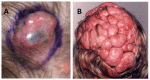
Cylindromas are well-circumscribed, smooth, pale pink nodular tumors, often with arborizing vessels visible. The tumors are slow growing and vary in size from a few millimeters to >5 mm (see Figure 1a). The finding of mixed cylindroma and spiradenoma histology within a single tumor is common in CYLD cutaneous syndrome, and the two terms have been used to describe variants of the same tumor [Rajan et al 2011a].

Figure 1. A.

Figure 1

A. Cylindroma demonstrating a well-circumscribed pink nodular lesion with arborizing blood vessels visible on the surface B. Confluent scalp cylindromas in a severely affected individual with CYLD cutaneous syndrome

Confluent scalp tumors. In severe cases, tumors may cover most of the scalp, referred to as confluent scalp tumors (see Figure 1b).

  • Early surgical intervention may prevent or delay confluent scalp tumors.
  • One study of two families reported that confluent scalp tumors affected up to one in four individuals with a heterozygous germline pathogenic CYLD variant [Rajan et al 2009a].

Pulmonary cylindromas. Single or multiple pulmonary cylindromas that originate from the skin have been described in three individuals with CYLD cutaneous syndrome [Vernon et al 1988, Brown et al 2018b]. In these cases, there is no history of a primary malignant cutaneous cylindroma in the skin, lymph node disease is absent, and pulmonary histology is benign, leading them to be categorized as "benign" metastases.

  • One individual presented at age 64 with breathlessness on exertion and was found to have multiple pulmonary tumors. This individual required serial monitoring with pulmonary imaging, and received endocopic laser ablation to maintain large airway patency.
  • A second individual had four asymptomatic pulmonary tumors discovered incidentally on a chest radiograph at age 80 years. The tumors were histologically confirmed on autopsy.
  • Whole-exome and genome sequencing have shown pulmonary cylindromas to harbor an additional pathogenic variant in AKT1 and a UV mutation signature confirming cutaneous origin [Davies et al 2019].
  • The true prevalence of pulmonary cylindromas in individuals with CYLD cutaneous syndrome is not known, as prospective radiologic imaging studies of large numbers of affected individuals have yet to be performed.
  • Currently, there are no routine surveillance imaging guidelines that can be recommended; however clinicians who care for individuals with CYLD cutaneous syndrome need to be aware that these tumors do arise and may need to be monitored to determine rate of growth, and that surgical interventions may be necessary to ablate pulmonary tumors that threaten large airway patency.

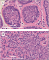
Histopathology. The histologic appearance of well-circumscribed cylindrical nests of basaloid cells in the dermis led to the term "cylindroma." Each cluster consists of darker, basophilic cells at the periphery, and larger pale cells centrally and is surrounded by a thick, hyaline membrane consisting of extracellular matrix proteins (including collagen IV and VII and laminin-332) in the basement membrane of the skin (see Figure 2a).

Figure 2. A.

Figure 2

A. Histopathology of cylindroma B. Histopathology of spiradenoma

Spiradenomas

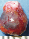
Spiradenomas are nodular tumors that are often blue/black in color. They tend to be painful and can grow up to 10 cm in diameter (see Figure 3).

Figure 3.

Figure 3.

Spiradenoma: a well-circumscribed nodular lesion (excision specimen) with characteristic blue/black appearance Rajan & Ashworth [2015]; republished with permission of author.

Histopathology. Spiradenomas are relatively disorganized histologically when compared to cylindromas and consist of sheets of epithelial cells associated with a lymphocytic infiltrate (mixed T and B cells). Some affected individuals present with histologic features of both cylindroma and spiradenoma within a single tumor specimen, giving rise to the term spiradenocylindroma. In addition, there is evidence that histologically organized cylindroma and histologically disorganized spiradenoma represent extremes of a spectrum of histophenotype of the same tumor [Rajan et al 2011a] (see Figure 2b).

Trichoepitheliomas

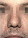
Trichoepitheliomas are skin-colored papules or firm nodules, mainly found on the central face (see Figure 4). They are often symmetrically distributed and usually no more than 2-5 mm across.

Figure 4.

Figure 4.

Trichoepithelioma: small skin-colored papules with a predilection for the central face Rajan & Ashworth [2015]; republished with permission of author.

Histopathology. Trichepitheliomas demonstrate clusters of basaloid germinative cells with keratinizing cystic spaces and superficial follicular differentiation surrounded by a fibrocytic stroma. Intra-stromal clefts and mesenchymal papillary buds may be seen.

Other Findings

Salivary lesions. Affected individuals are also at risk of developing tumors of the salivary glands, typically membranous basal cell adenoma (MBCA) [Jungehülsing et al 1999] usually after age 40 years.

  • These tumors typically present as a lump in the parotid gland, may be bilateral, and warrant a biopsy to confirm the diagnosis.
  • MBCA is a benign entity which may be managed surgically, but recurs in up to 25% of affected individuals [Zarbo 2002] (see also Malignant transformation).

Malignant transformation has been (rarely) reported in preexisting spiradenomas, cylindromas, spiradenocylindromas, and MBCA [Hyman et al 1988, Kazakov 2016]. Malignant tumors tend to be aggressive carcinomas with frequent local infiltrative growth or metastases.

  • Transcalvarial invasion is uncommon, but has been observed [Serracino & Kleinschmidt-Demasters 2013].
  • Malignant metastases to bone, lung, and liver have been reported [Gerretsen et al 1993].
  • Death from metastatic disease has been described in affected individuals in the early fifth decade [Kazakov et al 2009].
  • Malignant histopathology. Four diverse histologic patterns of malignant cylindroma or spiradenoma have been described [Kazakov et al 2009], including:
    • Salivary gland type basal cell adenocarcinoma-like pattern, low-grade
    • Salivary gland type basal cell adenocarcinoma-like pattern, high-grade
    • Invasive adenocarcinoma, not otherwise specified
    • Sarcomatoid (metaplastic) carcinoma

Other skin lesions and malignancies

  • Affected individuals may also develop small milia cysts on the skin of the face [Bajwa et al 2018].
  • Vulval cysts consisting histologically of multiple epidermal inclusion cysts and milia were reported in one affected female [Dubois et al 2017].
  • Squamous cell carcinoma [Ganguly et al 2012, Ma et al 2016] and follicular squamous cell carcinoma [Dubois et al 2018] arising in individuals with CYLD cutaneous syndrome have been described in isolated reports.
  • Basal cell carcinoma (BCC) is the most common human cancer, and coexistence in individuals with CYLD cutaneous syndrome may be coincidental or related to overlap in the appearance of each under the microscope. However, in a recent study of a large South American family with CYLD cutaneous syndrome, BCC was a reported in 25% of affected family members from this kindred [Arruda et al 2020].

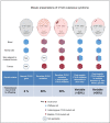
Segmental disease as a result of mosaic pathogenic CYLD variants should be considered when individuals develop unilateral clusters of any cylindromas, spiradenomas, or trichoepitheliomas [Arefi et al 2019]. Findings may include skin lesions arranged in a linear fashion [Furuichi et al 2012] following embryologic skin development lines (lines of Blaschko). The presentation of unilateral clusters may reflect either a pathogenic CYLD variant in the skin alone (late postzygotic mosaicism) or a pathogenic variant in both the blood and the skin (early postzygotic mosaicism) (see Figure 5) [Arefi et al 2019].

Figure 5.

Figure 5.

Mosaic presentations of CYLD cutaneous syndrome Arefi et al [2019]; published under Creative Commons license.

Genotype-Phenotype Correlations

No convincing genotype-phenotype correlations have been identified. There is a suggestion that individuals with pathogenic missense variants may have a milder phenotype; however, as missense variants constitute a minority of pathogenic variants in affected individuals (most have pathogenic truncating variants, which can also result in a mild phenotype), further studies are needed to investigate this hypothesis [Nagy et al 2015].

Nomenclature

Historically, descriptive names including Brooke-Spiegler syndrome (BSS), familial cylindromatosis (FC), and multiple familial trichoepithelioma (MFT) were assigned on the basis of the predominant tumor type and location. These conditions are now recognized to constitute a clinical spectrum and individuals with the clinical phenotypes of BSS, FC, and MFT can all present in a single family. The lack of prognostication offered by these historical labels favors the use of CYLD cutaneous syndrome as a diagnostic term for individuals with this single-gene disorder.

Outdated terms previously used in the literature to refer to CYLD cutaneous syndrome include the following:

  • Ancell-Spiegler cylindromas
  • Dermal eccrine cylindroma
  • Turban tumor syndrome (now denoted as confluent scalp tumors)

Prevalence

The true prevalence of CYLD cutaneous syndrome is unknown but may be in the order of more than 1:100,000 population [Rajan & Ashworth 2015].

Differential Diagnosis

Disorders with multiple facial papules in the differential diagnosis of CYLD cutaneous syndrome (CCS) are summarized in Table 3.

Table 3.

Disorders with Multiple Facial Papules in the Differential Diagnosis of CYLD Cutaneous Syndrome (CCS)

Gene(s)Disorder 1Distinguishing Histologic Features in Differential DisorderComment
FLCN Birt-Hogg-Dubé syndrome Fibrofolliculomas
NF1 Neurofibromatosis 1 (NF1)NeurofibromasBoth NF1 & CCS are assoc w/lesions on the torso
PTEN Cowden syndrome
(See PTEN Hamartoma Tumor Syndrome.)
Trichilemmomas
TSC1
TSC2
Tuberous sclerosis complex Angiofibromas
HR Marie Unna hypotrichosis 1 (MUHH1)
(OMIM 146550)
Trichoepitheliomas 2Severe hair breakage/loss & absence of cylindromas in MUHH1 further distinguishes MUHH1 from CCS.
PTCH1 Nevoid basal cell carcinoma syndrome Basal cell neviMacrocephaly, broad nasal root & jaw cysts
UnknownMultiple syringomas
(OMIM 186600)
Syringomas
1.

All of the disorders listed are inherited in an autosomal dominant manner.

2.

Pilar (trichilemmal) cysts. Pilar cysts are common keratin-filled cysts that often affect the scalp in multiple numbers, either sporadically or inherited as an autosomal dominant trait associated with heterozygous variants in PLCD1 [Hörer et al 2019]. They may mimic scalp cylindromas, but clinically, the skin overlying these scalp lesions is normal, while in cylindromas there is frequently loss of overlying hair and thinning of the skin, giving it a pink translucent appearance.

Management

Evaluations Following Initial Diagnosis

To establish the extent of disease and needs in an individual diagnosed with CYLD cutaneous syndrome (CCS), the evaluations summarized in Table 4 (if not performed as part of the evaluation that led to the diagnosis) are recommended.

Table 4.

Recommended Evaluations Following Initial Diagnosis in Individuals with CYLD Cutaneous Syndrome

System/ConcernEvaluationComment
Skin Skin exam by dermatologist
  • Full skin exam incl skin of the genitalia
  • Painful tumors should be identified & prioritized for excision (see Table 5).
  • Education about signs & symptoms of malignant transformation 1
Histologic examOf tumors that are rapidly growing, have a distinct appearance, or are ulcerated or bleeding
Imaging, if clinically indicated
  • Routine imaging not currently recommended
  • If malignant transformation is suspected in a scalp tumor, consider radiologic imaging (preferably MRI), given the possibility of intracranial invasion.
  • Little evidence is available as to when staging imaging should be considered for malignant tumors; consult specialist skin cancer multidisciplinary team. 2
Ears/Hearing Eval of external auditory canals w/otoscope
  • To screen for tumors that occlude the external auditory canal
  • When present, clinical assessment for conductive hearing loss may be considered.
Oral Clinical exam of parotid glandsTo screen for salivary lesions
Respiratory Assessment for signs of respiratory compromise in those w/new onset of shortness of breath, cough, or stridorIf present, radiologic imaging may be necessary to evaluate for pulmonary lesions.
Genetic
counseling
Consultation w/clinical geneticist &/or genetic counselorTo incl genetic counseling & cascade testing where needed
1.

Including tumors that are rapidly growing, bleeding, ulcerating or appear different than an affected individual's usual tumors.

2.

This team typically includes a dermatologist, plastic surgeon, radiologist, oncologist and pathologist, all of whom can guide a consensus decision making process.

Treatment of Manifestations

Table 5.

Treatment of Manifestations in Individuals with CYLD Cutaneous Syndrome

Manifestation/
Concern
TreatmentConsiderations/Other
Cylindroma, spiradenoma, trichoepithelioma Removal of tumors by conventional surgery
  • Repeated surgical procedures to ↓ tumor burden typically required 1,2
  • "Scalp-sparing" strategies incl early primary excision, tumor enucleation, & excision followed by secondary intention healing techniques recommended; 3 avoid removing large areas of scalp.
  • Complete scalp excision to be used only when no feasible alternatives 4
HyfrecationFor selected small tumors, such as trichoepithelioma on the nasolabial skin 5
Laser
  • Ablative laser resurfacing of smaller lesions (i.e., perinasal trichoepitheliomas) can yield good cosmetic results. 6
  • However, the advantage of laser treatment for large cylindromas & spiradenomas over standard excision is not clear. Laser treatment also precludes histologic assessment.
Mohs micrographic surgery 7
  • Technique allows dermatologic surgeon to track invasive tumor cells & confirm histologic clearance before closing the skin defect. 8
  • Mohs may be of limited benefit in CCS, as it may be difficult to obtain tumor-negative margins, leading to large defects.
Malignant tumors Multidisciplinary team input required to develop management plan
  • At least 8 different types of malignant tumors are seen in affected persons; w/the exception of BCC, these would be considered rare cancers & require appropriate eval following histopathologic assessment (see Clinical Characteristics).
  • Each case may need to be assessed & staged; decision should be made by a skin cancer multidisciplinary team w/support from dermatologists, oncologists, pathologists, plastic surgeons, & radiologists.

BCC = basal cell carcinoma; CCS = CYLD cutaneous syndrome

1.

Tumors progressively grow over time such that early treatment may reduce the need for extensive surgical procedures and also allow for the use of local anesthetic.

2.

Benign lesions removed for cosmetic reasons should have narrow margins to leave as much normal healthy tissue as possible.

3.
4.

Complete scalp excision is not curative. Often tumors develop from follicles within the graft, and individuals can have significant tumors at other sites, such as on the trunk.

5.

Histologic assessment of a representative lesion may be useful prior to ablative procedures. If there is a clinical suspicion of basal cell carcinoma, a biopsy should be performed prior to initiating ablative procedures.

6.

Repeated treatments are often needed and may not be cost effective.

7.

This technique has been used to treat sporadic cylindroma and spiradenoma where recurrence has occurred after primary surgical excision.

8.

Mohs may be best reserved for sporadic cases.

Prevention of Primary Manifestations

The role of UV light in the pathogenesis of the tumors seen in CYLD cutaneous syndrome is unclear. As with all skin cancer predisposition syndromes, appropriate precautions against UV-related skin damage are recommended [Davies et al 2019].

Surveillance

It is recommended that individuals with CYLD cutaneous syndrome undergo at least annual full skin examination by a dermatologist, with some affected individuals requiring skin review every three to four months.

  • An assessment of tumor burden and rate of new tumor development can be made, and existing tumors can also be monitored for any signs of malignant transformation.
  • Annual monitoring will guide the frequency and interval of surgical procedures.
  • Between appointments, affected individuals should be asked to report growing, ulcerated, or bleeding tumors or tumors that appear different from existing lesions so that they can be assessed to determine if urgent excision is warranted.

Agents/Circumstances to Avoid

Radiotherapy should be avoided as it causes DNA damage and may result in further tumor formation or malignant transformation of existing lesions [Crain & Helwig 1961, Rajan & Ashworth 2015].

Evaluation of Relatives at Risk

It is appropriate to clarify the genetic status of apparently asymptomatic older and younger at-risk relatives of an affected individual in order to identify as early as possible those who would benefit from prompt initiation of treatment and preventive measures.

See Genetic Counseling for issues related to testing of at-risk relatives for genetic counseling purposes.

Therapies Under Investigation

Treatment for CYLD cutaneous syndrome is largely surgical. The first placebo-controlled early-phase trial of a topical targeted kinase inhibitor (tropomyosin receptor kinase) showed short-term safety [Danilenko et al 2018]; a dose escalation study is needed to determine efficacy.

It is important to recognize that tumors in CYLD cutaneous syndrome lack a curative medical therapy, and any treatments will likely need to be repeated over the affected individual's lifetime. Isolated case reports of topical or intralesional therapeutic interventions must be interpreted with caution as all have some or all of the following limitations:

  • They are not placebo controlled.
  • No objective measures were used to assess improvement.
  • Only short-term follow-up data are presented.
  • No long-term safety data regarding the repeated use of these agents in treatment of CYLD cutaneous syndrome are available.

Search ClinicalTrials.gov in the US and EU Clinical Trials Register in Europe for access to information on clinical studies for a wide range of diseases and conditions.

Genetic Counseling

Genetic counseling is the process of providing individuals and families with information on the nature, mode(s) of inheritance, and implications of genetic disorders to help them make informed medical and personal decisions. The following section deals with genetic risk assessment and the use of family history and genetic testing to clarify genetic status for family members; it is not meant to address all personal, cultural, or ethical issues that may arise or to substitute for consultation with a genetics professional. —ED.

Mode of Inheritance

CYLD cutaneous syndrome (CCS) is inherited in an autosomal dominant manner.

Risk to Family Members

Parents of a proband

  • Most individuals diagnosed with CYLD cutaneous syndrome have an affected parent. Because the degree of severity can vary within families, a mildly affected parent may have a more severely affected child and vice versa.
  • Some individuals diagnosed with CYLD cutaneous syndrome have the disorder as the result of a de novo CYLD pathogenic variant. Simplex cases (i.e., a single occurrence in a family) have not been evaluated sufficiently to determine if the pathogenic variant occurred de novo, and thus the proportion of CYLD cutaneous syndrome caused by a de novo pathogenic variant is unknown.
  • Molecular genetic testing is recommended for the parents of a proband with an apparent de novo pathogenic variant.
  • If the pathogenic variant found in the proband cannot be detected in the leukocyte DNA of either parent, possible explanations include a de novo pathogenic variant in the proband or somatic and/or germline mosaicism in a parent.* One instance of a proband inheriting a pathogenic variant from a parent with germline mosaicism has been reported [Arefi et al 2019].
    * Misattributed parentage can also be explored as an alternative explanation for an apparent de novo pathogenic variant.
  • The family history of some individuals diagnosed with CYLD cutaneous syndrome may appear to be negative because of failure to recognize the disorder in family members, a mild phenotype, early death of the parent before the onset of symptoms, or late onset of the disease in the affected parent. Therefore, an apparently negative family history cannot be confirmed unless molecular genetic testing has been performed on the parents of the proband.
    Note: If the parent is the individual in whom the pathogenic variant first occurred, the parent may have somatic mosaicism for the variant and may be minimally affected. Such individuals may present with a unilateral cluster of cylindromas, spiradenomas, or trichoepitheliomas

Sibs of a proband. The risk to the sibs of the proband depends on the clinical/genetic status of the proband's parents:

  • If a parent of the proband is affected and/or is known to be heterozygous for the pathogenic variant identified in the proband, the risk to the sibs is 50%. Although the penetrance of CYLD cutaneous syndrome approaches 100%, clinical severity as well as the pattern of presentation of the tumors (e.g., on the scalp vs the face) and tumor types may vary among heterozygous sibs.
  • If the proband has a known CYLD pathogenic variant that cannot be detected in the leukocyte DNA of either parent and both parents are clinically unaffected, the recurrence risk to sibs is slightly greater than that of the general population because of the possibility of parental germline mosaicism.
  • If clinical evaluation of a parent suggests a mosaic presentation of CYLD cutaneous syndrome and molecular genetic testing is consistent with postzygotic mutation of CYLD in the parent, the risk to sibs is variable but is presumed to be less than 50% [Arefi et al 2019] (see Figure 5).

Offspring of a proband. Each child of an individual with a heterozygous CYLD pathogenic variant has a 50% chance of inheriting the pathogenic variant. Because intrafamilial clinical variability is observed in CYLD cutaneous syndrome, offspring who inherit a CYLD pathogenic variant may be more or less severely affected than the transmitting parent.

Other family members. The risk to other family members depends on the status of the proband's parents: if a parent has a CYLD pathogenic variant, the parent's family members may be at risk.

Related Genetic Counseling Issues

See Management, Evaluation of Relatives at Risk for information on evaluating at-risk relatives for the purpose of early diagnosis and treatment.

Predictive testing for at-risk asymptomatic adult family members requires prior identification of the CYLD pathogenic variant in the family.

Family planning

  • The optimal time for determination of genetic risk and discussion of the availability of prenatal/preimplantation genetic testing is before pregnancy.
  • It is appropriate to offer genetic counseling (including discussion of potential risks to offspring and reproductive options) to young adults who are affected or at risk.

DNA banking. Because it is likely that testing methodology and our understanding of genes, pathogenic mechanisms, and diseases will improve in the future, consideration should be given to banking DNA from probands in whom a molecular diagnosis has not been confirmed (i.e., the causative pathogenic mechanism is unknown).

Prenatal Testing and Preimplantation Genetic Testing

Once the CYLD pathogenic variant has been identified in an affected family member, prenatal testing for a pregnancy at increased risk and preimplantation genetic testing are possible.

Differences in perspective may exist among medical professionals and within families regarding the use of prenatal testing. While most centers would consider use of prenatal testing to be a personal decision, discussion of these issues may be helpful.

Resources

GeneReviews staff has selected the following disease-specific and/or umbrella support organizations and/or registries for the benefit of individuals with this disorder and their families. GeneReviews is not responsible for the information provided by other organizations. For information on selection criteria, click here.

Molecular Genetics

Information in the Molecular Genetics and OMIM tables may differ from that elsewhere in the GeneReview: tables may contain more recent information. —ED.

Table A.

CYLD Cutaneous Syndrome: Genes and Databases

GeneChromosome LocusProteinLocus-Specific DatabasesHGMDClinVar
CYLD 16q12​.1 Ubiquitin carboxyl-terminal hydrolase CYLD CYLD database CYLD CYLD

Data are compiled from the following standard references: gene from HGNC; chromosome locus from OMIM; protein from UniProt. For a description of databases (Locus Specific, HGMD, ClinVar) to which links are provided, click here.

Table B.

OMIM Entries for CYLD Cutaneous Syndrome (View All in OMIM)

132700CYLINDROMATOSIS, FAMILIAL
601606TRICHOEPITHELIOMA, MULTIPLE FAMILIAL, 1
605018CYLD LYSINE-63 DEUBIQUITINASE; CYLD
605041BROOKE-SPIEGLER SYNDROME; BRSS

Molecular Pathogenesis

CYLD encodes the tumor suppressor ubiquitin carboxyl-terminal hydrolase, an enzyme involved in the deubiquitination of "Lys-63" (K63) [Brummelkamp et al 2003, Trompouki et al 2003] and "Met-1" linked ubiquitin chains [Draber et al 2015, Kupka et al 2016]. CYLD negatively regulates NF-κB by removing Lys-63 linked ubiquitin chains from TRAF2, TRAF6, and NEMO, thus tempering the activity of NF-κB. In addition, CYLD deubiquitinates other substrates within the NF-κB signaling pathway, such as BCL3 and TAK1. In CYLD cutaneous syndrome tumors, CYLD is lost, and the consequently increased NF-κB signaling may give tumor cells a survival advantage via proliferation and resistance to apoptosis.

CYLD also negatively regulates a range of signaling pathways that are important in inflammation and cancer, including JNK, Wnt, TGFB1, and Notch [Rajan & Ashworth 2015]. Transcriptome studies of cylindromas and spiradenomas demonstrate evidence of increased NF-κB and Wnt signaling [Rajan et al 2011a, Rajan et al 2011b].

Mechanism of disease causation. As is the case for many tumor predisposition syndromes, CYLD cutaneous syndrome occurs through a loss-of-function mechanism that requires a somatic "second hit" for disease manifestation. In CYLD cutaneous syndrome tumors, loss of the remaining normal allele is frequently seen (loss of heterozygosity), either by reduplication of the entire arm of 16q harboring the germline pathogenic variant, or by a second pathogenic variant in the remaining normal allele. Recent work has suggested that recurrent pathogenic variants in DNMT3A and BCOR may also occur in addition to biallelic CYLD loss, suggesting that epigenetic dysregulation may contribute to tumor pathogenesis [Davies et al 2019].

CYLD-specific laboratory technical considerations. For mosaic cases, it may be necessary to study tumor tissue from multiple skin samples. This can be technically challenging on FFPE tissue; if possible, fresh skin biopsy tissue should be used for DNA extraction.

Cancer and Benign Tumors

Somatic CYLD pathogenic variants have been found in sporadic cylindromas and spiradenomas [Bignell et al 2000]. They have also been found in salivary gland tumors such as basal cell adenocarcinoma [Rito et al 2018] and adenoid cystic carcinoma [Stephens et al 2013]. In hematologic malignancies, there are reports of CYLD deletion playing a role in myeloma, where a recurrent contiguous deletion involving CYLD and WWOX is noted to be associated with a worse prognostic outcome [Jenner et al 2007]. CYLD pathogenic variants have been reported in an isolated case of Hodgkin lymphoma [Schmidt et al 2010].

References

Literature Cited

  • Arefi M, Wilson V, Muthiah S, Zwolinski S, Bajwa D, Brennan P, Blasdale K, Bourn D, Burn J, Santibanez-Koref M, Rajan N. Diverse presentations of cutaneous mosaicism occur in CYLD cutaneous syndrome and may result in parent-to-child transmission. J Am Acad Dermatol. 2019;81:1300–7. [PMC free article: PMC6878220] [PubMed: 31085270]
  • Arruda AP, Cardoso-Dos-Santos AC, Mariath LM, Feira MF, Kowalski TW, Bezerra KRF, da Silva LACT, Ribeiro EM, Schuler-Faccini L. 2019. A large family with CYLD cutaneous syndrome: medical genetics at the community level. J Community Genet. 2020;11:279–84. [PMC free article: PMC7295879] [PubMed: 31792733]
  • Bajwa DS, Nasr B, Carmichael AJ, Rajan N. Milia: a useful clinical marker of CYLD mutation carrier status. Clin Exp Dermatol. 2018;43:193–5. [PubMed: 29023940]
  • Bignell GR, Warren W, Seal S, Takahashi M, Rapley E, Barfoot R, Green H, Brown C, Biggs PJ, Lakhani SR, Jones C, Hansen J, Blair E, Hofmann B, Siebert R, Turner G, Evans DG, Schrander-Stumpel C, Beemer FA, van Den Ouweland A, Halley D, Delpech B, Cleveland MG, Leigh I, Leisti J, Rasmussen S. Identification of the familial cylindromatosis tumor-suppressor gene. Nat Genet. 2000;25:160–5. [PubMed: 10835629]
  • Brass D, Rajan N, Langtry J. Enucleation of cylindromas in brooke-spiegler syndrome: a novel surgical technique. Dermatol Surg. 2014;2014;40:1438–9. [PMC free article: PMC6082342] [PubMed: 25361203]
  • Brown SM, Arefi M, Stones R, Loo PS, Barnard S, Bloxham C, Stefanos N, Langtry JAA, Worthy S, Calonje E, Husain A, Rajan N. Inherited pulmonary cylindromas: extending the phenotype of CYLD mutation carriers. Br J Dermatol. 2018a;179:662–8. [PMC free article: PMC6175122] [PubMed: 29569226]
  • Brown S, Worthy SA, Langtry JA, Rajan N. Tracking tumor kinetics in patients with germline CYLD mutations. J Am Acad Dermatol. 2018b;79:949–51. [PMC free article: PMC6207503] [PubMed: 29660420]
  • Brummelkamp TR, Nijman SM, Dirac AM, Bernards R. Loss of the cylindromatosis tumor suppressor inhibits apoptosis by activating NF-kappaB. Nature. 2003;2003;424:797–801. [PubMed: 12917690]
  • Crain RC, Helwig EB. Dermal cylindroma (dermal eccrine cylindroma). Am J Clin Pathol. 1961;35:504–15. [PubMed: 13696220]
  • Danilenko M, Stamp E, Stocken DD, Husain A, Zangarini M, Cranston A, Stones R, Sinclair N, Hodgson K, Bowett SA, Roblin D, Traversa S, Plummer R, Veal G, Langtry JAA, Ashworth A, Burn J, Rajan N. Targeting tropomyosin receptor kinase in cutaneous CYLD defective tumors with pegcantratinib: the TRAC randomized clinical trial. JAMA Dermatol. 2018;154:913–21. [PMC free article: PMC6128505] [PubMed: 29955768]
  • Davies HR, Hodgson K, Schwalbe E, Coxhead J, Sinclair N, Zou X, Cockell S, Husain A, Nik-Zainal S, Rajan N. Epigenetic modifiers DNMT3A and BCOR are recurrently mutated in CYLD cutaneous syndrome. Nat Commun. 2019;10:4717. [PMC free article: PMC6797807] [PubMed: 31624251]
  • Draber P, Kupka S, Reichert M, Draberova H, Lafont E, De Miguel D, Spilgies L, Surinova S, Taraborrelli L, Hartwig T, Rieser E, Martino L, Rittinger K, Walczak H. LUBAC-recruited CYLD and A20 regulate gene activation and cell death by exerting opposing effects on linear ubiquitin in signaling complexes. Cell Rep. 2015;13:2258–72. [PMC free article: PMC4688036] [PubMed: 26670046]
  • Dubois A, Alonso-Sanchez A, Bajaj V, Husain A, Rajan N. Multiple facial trichoepitheliomas and vulval cysts. JAMA Dermatology. 2017;153:826–8. [PMC free article: PMC5817598] [PubMed: 28423152]
  • Dubois A, Mestre T, Oliphant T, Husain A, Rajan N. Squamous cell carcinoma and multiple familial trichoepitheliomas: a recurrent association. Acta Derm Venereol. 2018;98:910–11. [PubMed: 29972217]
  • Dubois A, Wilson V, Bourn D, Rajan N. CYLD genetic testing for Brooke-Spiegler syndrome, familial cylindromatosis and multiple familial trichoepitheliomas. PLoS Curr. 2015:7. [PMC free article: PMC4339271] [PubMed: 25737804]
  • ESHG. Abstracts from the 51st European Society of Human Genetics Conference: posters. Eur J Hum Genet. 2019;27:1–688.
  • Furuichi M, Makino T, Yamakoshi T, Matsui K, Shimizu T. Blaschkoid distribution of cylindromas in a germline CYLD mutation carrier. Br J Dermatol. 2012;166:1376–8. [PubMed: 22296260]
  • Ganguly S, Jaykar KC, Banerjee PK, Kumar R, Ahmed N. Multiple familial trichoepitheliomas in association with squamous cell carcinoma. Indian Dermatol Online J. 2012;3:151–3. [PMC free article: PMC3481869] [PubMed: 23130296]
  • Gerretsen AL, Van Der Putte SC, Deenstra W, Van Vloten WA. Cutaneous cylindroma with malignant transformation. Cancer. 1993;1993;72:1618–23. [PubMed: 7688655]
  • Grossmann P, Vanecek T, Steiner P, Kacerovska D, Spagnolo DV, Cribier B, Rose C, Vazmitel M, Carlson JA, Emberger M, Martinek P, Pearce RL, Pearn J, Michal M, Kazakov DV. Novel and recurrent germline and somatic mutations in a cohort of 67 patients from 48 families with Brooke-Spiegler syndrome including the phenotypic variant of multiple familial trichoepitheliomas and correlation with the histopathologic findings in 379 biopsy specimens. Am J Dermatopathol. 2013;35:34–44. [PubMed: 23249834]
  • Hörer S, Marrakchi S, Radner FPW, Zolles G, Heinz L, Eichmann TO, Has C, Salavei P, Mahfoudh N, Turki H, Zimmer AD, Fischer J. A monoallelic two-hit mechanism in PLCD1 explains the genetic pathogenesis of hereditary trichilemmal cyst formation. J Invest Dermatol. 2019;139:2154–63.e5. [PubMed: 31082376]
  • Huang Y, Cai C, Ren L, Cui C, Zhang X, Liu W. Marie Unna hereditary hypotrichosis accompanied by multiple familial trichoepithelioma in a Chinese family. J Dermatol. 2019;46:413–17. [PubMed: 30809827]
  • Hyman BA, Scheithauer BW, Weiland LH, Irons GB. Membranous basal cell adenoma of the parotid gland. Malignant transformation in a patient with multiple dermal cylindromas. Arch Pathol Lab Med. 1988;112:209–11. [PubMed: 2827601]
  • Jenner MW, Leone PE, Walker BA, Ross FM, Johnson DC, Gonzalez D, Chiecchio L, Dachs Cabanas E, Dagrada GP, Nightingale M, Protheroe RK, Stockley D, Else M, Dickens NJ, Cross NC, Davies FE, Morgan GJ. Gene mapping and expression analysis of 16q loss of heterozygosity identifies WWOX and CYLD as being important in determining clinical outcome in multiple myeloma. Blood. 2007;110:3291–300. [PubMed: 17609426]
  • Jungehülsing M, Wagner M, Damm M. Turban tumor with involvement of the parotid gland. J Laryngol Otol. 1999;113:779–83. [PubMed: 10748863]
  • Kazakov DV, Zelger B, Rütten A, Vazmitel M, Spagnolo DV, Kacerovska D, Vanecek T, Grossmann P, Sima R, Grayson W, Calonje E, Koren J, Mukensnabl P, Danis D, Michal M. Morphologic diversity of malignant neoplasms arising in preexisting spiradenoma, cylindroma, and spiradenocylindroma based on the study of 24 cases, sporadic or occurring in the setting of Brooke-Spiegler syndrome. Am J Surg Pathol. 2009;33:705–19. [PubMed: 19194280]
  • Kazakov DV. Brooke-Spiegler syndrome and phenotypic variants: an update. Head Neck Pathol. 2016;2016;10:125–30. [PMC free article: PMC4838966] [PubMed: 26971504]
  • Kupka S, De Miguel D, Draber P, Martino L, Surinova S, Rittinger K, Walczak H. SPATA2-mediated binding of CYLD to HOIP enables CYLD recruitment to signaling complexes. Cell Rep. 2016;16:2271–80. [PMC free article: PMC5009064] [PubMed: 27545878]
  • Ma H, Feng S, Pei W, Jin F. Twelve years' observation of multiple familial trichoepithelioma with squamous carcinoma. Indian J Dermatol. 2016;2016;61:348. [PMC free article: PMC4885207] [PubMed: 27293274]
  • Nagy N, Farkas K, Kemény L, Széll M. Phenotype–genotype correlations for clinical variants caused by CYLD mutations. Eur J Med Genet. 2015;2015;58:271–8. [PubMed: 25782638]
  • Nagy N, Rajan N, Farkas K, Kinyó A, Kemény L, Széll M. A mutational hotspot in CYLD causing cylindromas: a comparison of phenotypes arising in different genetic backgrounds. Acta Derm Venereol. 2013;93:743–5. [PMC free article: PMC6166779] [PubMed: 23584127]
  • Rajan N, Ashworth A. Inherited cylindromas: lessons from a rare tumor. Lancet Oncol. 2015;16:e460–9. [PubMed: 26370355]
  • Rajan N, Burn J, Langtry J, Sieber-Blum M, Lord CJ, Ashworth A. Transition from cylindroma to spiradenoma in CYLD-defective tumors is associated with reduced DKK2 expression. J Pathol. 2011a;224:309–21. [PubMed: 21598248]
  • Rajan N, Elliott R, Clewes O, Mackay A, Reis-Filho JS, Burn J, Langtry J, Sieber-Blum M, Lord CJ, Ashworth A. Dysregulated TRK signalling is a therapeutic target in CYLD defective tumors. Oncogene. 2011b;30:4243–60. [PMC free article: PMC3175103] [PubMed: 21552290]
  • Rajan N, Langtry JAA, Ashworth A, Roberts C, Chapman P, Burn J, Trainer AH. Tumor mapping in 2 large multigenerational families with CYLD mutations: implications for disease management and tumor induction. Arch Dermatol. 2009a;145:1277–84. [PMC free article: PMC2935681] [PubMed: 19917957]
  • Rajan N, Trainer AH, Burn J, Langtry JAA. Familial cylindromatosis and brooke-spiegler syndrome: a review of current therapeutic approaches and the surgical challenges posed by two affected families. Dermatol Surg. 2009b;35:845–52. [PubMed: 19397670]
  • Rashid M, van der Horst M, Mentzel T, Butera F, Ferreira I, Pance A, Rütten A, Luzar B, Marusic Z, de Saint Aubain N, Ko JS, Billings SD, Chen S, Abi Daoud M, Hewinson J, Louzada S, Harms PW, Cerretelli G, Robles-Espinoza CD, Patel RM, van der Weyden L, Bakal C, Hornick JL, Arends MJ, Brenn T, Adams DJ. ALPK1 hotspot mutation as a driver of human spiradenoma and spiradenocarcinoma. Nat Commun. 2019;10:2213. [PMC free article: PMC6525246] [PubMed: 31101826]
  • Rito M, Mitani Y, Bell D, Mariano FV, Almalki ST, Pytynia KB, Fonseca I, El-Naggar AK. Frequent and differential mutations of the CYLD gene in basal cell salivary neoplasms: linkage to tumor development and progression. Mod Pathol. 2018;31:1064–72. [PubMed: 29463883]
  • Saggar S, Chernoff KA, Lodha S, Horev L, Kohl S, Honjo RS, Brandt HRC, Hartmann K, Celebi JT. CYLD mutations in familial skin appendage tumors. J Med Genet. 2008;45:298–302. [PubMed: 18234730]
  • Schmidt A, Schmitz R, Giefing M, Martin-Subero JI, Gesk S, Vater I, Massow A, Maggio E, Schneider M, Hansmann ML, Siebert R, Kuppers R. Rare occurrence of biallelic CYLD gene mutations in classical Hodgkin lymphoma. Genes Chromosomes Cancer. 2010;2010;49:803–9. [PubMed: 20607853]
  • Serra AD, Rubinstein D, Law C, Kramer J, Davis C. Plain radiographic and MR imaging appearances of familial dermal cylindroma. AJR Am J Roentgenol. 1996;167:615–16. [PubMed: 8751662]
  • Serracino HS, Kleinschmidt-Demasters BK. Skull invaders: when surgical pathology and neuropathology worlds collide. J Neuropathol Exp Neurol. 2013;72:600–13. [PubMed: 23771219]
  • Stephens PJ, Davies HR, Mitani Y, Van Loo P, Shlien A, Tarpey PS, Papaemmanuil E, Cheverton A, Bignell GR, Butler AP, Gamble J, Gamble S, Hardy C, Hinton J, Jia M, Jayakumar A, Jones D, Latimer C, McLaren S, McBride DJ, Menzies A, Mudie L, Maddison M, Raine K, Nik-Zainal S, O'Meara S, Teague JW, Varela I, Wedge DC, Whitmore I, Lippman SM, McDermott U, Stratton MR, Campbell PJ, El-Naggar AK, Futreal PA. Whole exome sequencing of adenoid cystic carcinoma. J Clin Invest. 2013;123:2965–8. [PMC free article: PMC3999050] [PubMed: 23778141]
  • Trompouki E, Hatzivassiliou E, Tsichritzis T, Farmer H, Ashworth A, Mosialos G. CYLD is a deubiquitinating enzyme that negatively regulates NF-kappaB activation by TNFR family members. Nature. 2003;424:793–6. [PubMed: 12917689]
  • Vanecek T, Halbhuber Z, Kacerovska D, Martinek P, Sedivcova M, Carr RA, Slouka D, Michal M, Kazakov DV. Large germline deletions of the CYLD gene in patients with Brooke-Spiegler syndrome and multiple familial trichoepithelioma. Am J Dermatopathol. 2014;36:868–74. [PubMed: 25347032]
  • Vernon HJ, Olsen EA, Vollmer RT. Autosomal dominant multiple cylindromas associated with solitary lung cylindroma. J Am Acad Dermatol. 1988;19:397–400. [PubMed: 2842382]
  • Zarbo RJ. Salivary gland neoplasia: a review for the practicing pathologist. Mod Pathol. 2002;15:298–323. [PubMed: 11904344]

Chapter Notes

Author Notes

Dr Neil Rajan, MD, PhD is a senior lecturer and honorary consultant dermatologist based in Newcastle, UK. He has received fellowships from the Wellcome Trust and the Medical Research Council that have supported his work on the molecular dissection of inherited cutaneous tumor syndromes. His basic science research program is coupled with the delivery of early-phase clinical trials in rare disease, an exemplar of which is the TRAC study in CYLD cutaneous syndrome, where he was chief investigator. By working in partnership with patients with rare skin disease, his work aims to discover oncogenic dependencies in skin tumors that can be targeted for therapeutic benefit.

Dr Rajan's web page

Acknowledgments

We would like to take the opportunity to thank the patients who have generously participated in research.

Revision History

  • 16 April 2020 (ma) Review posted live
  • 13 September 2019 (nr) Original submission
Copyright © 1993-2024, University of Washington, Seattle. GeneReviews is a registered trademark of the University of Washington, Seattle. All rights reserved.

GeneReviews® chapters are owned by the University of Washington. Permission is hereby granted to reproduce, distribute, and translate copies of content materials for noncommercial research purposes only, provided that (i) credit for source (http://www.genereviews.org/) and copyright (© 1993-2024 University of Washington) are included with each copy; (ii) a link to the original material is provided whenever the material is published elsewhere on the Web; and (iii) reproducers, distributors, and/or translators comply with the GeneReviews® Copyright Notice and Usage Disclaimer. No further modifications are allowed. For clarity, excerpts of GeneReviews chapters for use in lab reports and clinic notes are a permitted use.

For more information, see the GeneReviews® Copyright Notice and Usage Disclaimer.

For questions regarding permissions or whether a specified use is allowed, contact: ude.wu@tssamda.

Bookshelf ID: NBK555820PMID: 32298062

Views

Tests in GTR by Gene

Related information

  • OMIM
    Related OMIM records
  • PMC
    PubMed Central citations
  • PubMed
    Links to PubMed
  • Gene
    Locus Links

Similar articles in PubMed

See reviews...See all...

Recent Activity

Your browsing activity is empty.

Activity recording is turned off.

Turn recording back on

See more...