Glycosaminoglycans bind to many different classes of proteins mostly through electrostatic interactions between negatively charged sulfate groups and uronic acids and positively charged amino acids in the protein. This chapter focuses on examples of glycosaminoglycan (GAG)-binding proteins, methods for measuring GAG–protein interaction, and information about three-dimensional structures of the complexes.
GAG-BINDING PROTEINS ARE COMMON
In contrast to lectins, which tend to fall into evolutionarily conserved families (Chapters 28–37), GAG-binding proteins do not have common folds and instead appear to have evolved by convergent evolution. Several hundred GAG-binding proteins have been discovered, which make up the GAG-interactome and fall into the broad classes presented in Table . To a large extent, studies of the GAG-interactome have focused on protein interactions with heparin, a more highly sulfated, iduronic acid (IdoA)-rich form of heparan sulfate (HS; Chapter 17). This bias reflects, in part, the commercial availability of heparin and heparin-Sepharose, which are frequently used for fractionation studies, and the partially incorrect assumption that binding to heparin mimics binding to HS present on cell surfaces and in the extracellular matrix. There are also a large number of proteins known to interact with chondroitin sulfate (CS) and dermatan sulfate (DS) with comparable avidity and affinity; there are fewer examples of specific interactions with keratan sulfate (KS), but this may reflect fewer studies of KS. In some cases, CS may be the physiologically relevant ligand because CS predominates in many tissues. Determining the physiological relevance of these interactions is a major area of research.
Examples of various classes of glycosaminoglycan (GAG)-binding proteins
Interactions between GAGs and proteins can have profound physiological effects on processes such as hemostasis, lipid transport and absorption, cell growth and migration, and development. Binding to GAGs can result in immobilization of proteins at their sites of production or in the extracellular matrix for future mobilization, regulation of enzyme activity, binding of proteins to their receptors, protein oligomerization, and protection of proteins against degradation. Several viruses and bacteria also exploit GAGs expressed in the extracellular matrix and on cell surfaces as attachment factors. For example, SARS-CoV-2, the causative agent of the COVID-19 pandemic, enters through the respiratory tract and infects epithelial cells lining the airways by interactions of the viral spike protein with heparan sulfate. The ability of virions to attach to cell-surface heparan sulfate facilitates capture of the virus and transfer to proteinaceous receptors (e.g., host cell receptor angiotensin converting enzyme 2 in the case of SARS-CoV-2) as well as subsequent viral glycoprotein processing and infection. The interactions of GAG-binding proteins are often driven by a general complementarity of charge (e.g., thrombin–heparin interactions). In some cases, the interaction has been shown to depend on rare but specific sequences of modified sugars in the GAG chain (e.g., antithrombin binding).
METHODS FOR MEASURING GAG–PROTEIN BINDING
Numerous methods are available for analyzing GAG–protein interactions, and some provide a direct measurement of Kd values. A common method involves affinity fractionation of proteins on Sepharose columns containing covalently linked GAG chains, usually heparin. The bound proteins are eluted with different concentrations of sodium chloride, and the concentration required for elution is generally proportional to the Kd. High-affinity interactions require 0.5–2 m NaCl to displace bound ligand, which usually translates into Kd values of 10−7–10−9
m (determined under physiological salt concentrations by equilibrium binding). Proteins with lower affinity (10−5–10−7
m) often require only 0.2–0.5 m NaCl to elute. This method assumes that GAG–protein interactions are entirely ionic, which is not entirely correct. Nevertheless, it can provide an assessment of relative affinity, when comparing different GAG-binding proteins.
A number of more sophisticated methods are now in use that provide detailed thermodynamic data (ΔH [change in enthalpy], ΔS [change in entropy], ΔCp [change in molar heat capacity], etc.), kinetic data (association and dissociation rates), and high-resolution data on atomic contacts in GAG–protein interactions (Table ). Regardless of the technique one uses, it must be kept in mind that in vitro binding measurements are not likely to be the same as those when the protein binds to proteoglycans on the cell surface or in the extracellular matrix, where the density and variety of GAG-binding proteins, proteoglycans, and other interacting factors varies greatly. To determine the physiological relevance of the interaction, one should consider measuring binding under conditions that can lead to a biological response. For example, one can measure binding to cells with altered GAG composition (Chapter 49) or after treatment with specific lyases to remove GAG chains from the cell surface (Chapter 17) and then determine whether the same response occurs as observed in the presence of GAG chains. The interaction can then be studied more intensively using the in vitro assays described above.
Methods to measure glycosaminoglycan (GAG)–protein interaction
CONFORMATIONAL AND SEQUENCE CONSIDERATIONS
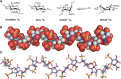
As mentioned above, most GAG-binding proteins interact with HS and/or heparin. The likely basis for this preference is greater sequence heterogeneity and more extensive and variable sulfation compared with other GAGs. The unusual conformational flexibility of iduronic acid, which is found in heparin, HS, and DS, also has a role in their ability to bind proteins. GAGs are linear helical structures, consisting of alternating residues of N-acetylglucosamine (GlcNAc) or N-acetylgalactosamine (GalNAc) with glucuronic acid (GlcA) or IdoA (with the exception of KS, which consists of alternating N-acetylglucosamine and galactose residues; Chapter 17). Inspection of heparin oligosaccharides containing highly modified domains ([GlcNS6S-IdoA2S]n) shows that the N-sulfo and 2-O-sulfo groups of each disaccharide repeat lie on opposite sides of the helix from the 6-O-sulfo and carboxyl groups (Figure ). Analysis of the conformation of individual sugars shows that N-acetylglucosamine and glucuronic acid residues assume a preferred conformation in solution, designated 4C1 (indicating that carbon 4 is above the plane defined by carbons 2, 3, and 5 and the ring oxygen, and that carbon 1 is below the plane; Chapter 2). In contrast, IdoA2S assumes the 1C4 or the 2S0 conformation (Figure ), which reorients the position of the sulfo substituents, thereby creating a different orientation of charged groups. In many cases when a protein binds to an HS chain, it induces a change in conformation of the IdoA2S residue resulting in a better fit and enhanced binding. IdoA2S residues have always been found in domains rich in N-sulfo and O-sulfo groups (for biosynthetic reasons; Chapter 17), which is also where proteins usually bind. Thus, the greater degree of conformational flexibility in these modified regions may explain why so many more proteins bind with high affinity to heparin, HS, and DS than to other GAGs. The presence of an N-acetyl group in an N-acetylglucosamine residue changes the preferred conformation of the neighboring IdoA residue, showing that even minor modifications can influence conformation and chain flexibility. Binding to GAGs that have a low degree of sulfation may require larger domains in the protein to interact with longer stretches of an oligosaccharide. Molecular dynamic simulations on large heparin oligosaccharides, even in the presence of proteins, are possible with recent advances in computer performance (Online Appendix 38A). Such simulations can be used to predict the conformational flexibility of different domains within the chain and, when combined with recent advances in protein–GAG docking, can provide additional insights into GAG–protein interactions.
Conformation of heparin oligosaccharides. (A) Glucosamine (GlcN) and glucuronic acid (GlcA) exist in the 4C1 conformation, whereas iduronic acid (IdoA) exists in equally energetic conformations designated 1C4 and 2S0. (B) Space-filling model of a heparin (more...)
HOW SPECIFIC ARE GAG–PROTEIN INTERACTIONS?
The discovery of multiple GAG-binding proteins led a number of investigators to examine whether there is a consensus amino acid sequence for GAG binding. In retrospect, this strategy was overly simplistic because it assumed that all GAG-binding proteins have a common evolutionary origin and would recognize the same oligosaccharide sequence within heparin or, at least, sequences that would share many common features. It is now known that the convergently evolved GAG-binding proteins interact with different oligosaccharide sequences. The binding sites in the protein always contain basic amino acids (lysine and arginine), whose positive charges presumably interact with the negatively charged sulfates and carboxylates of the GAG chains. However, the arrangement of these basic amino acids can be quite variable, consistent with the variable positioning of sulfo groups in the GAG partner. Selectivity is also a function of H-bonding and van der Waal interactions of amino acid residues with the oligosaccharide.
Most proteins are formed from α-helices, β-strands, and loops. Therefore, to engage a linear GAG chain electrostatically, the positively charged amino acid residues must align along the same side of the protein segment. α-Helices have periodicities of 3.4 residues per turn, which would require the basic residues to occur every third or fourth position along the helix to align with an oligosaccharide. In β-strands, the amino acid side chains alternate sides every other residue. Thus, to bind a GAG chain, the positively charged residues in a β-strand would be located quite differently than in an α-helix.
On the basis of the structure of several heparin-binding proteins that were available in 1991, Alan Cardin and Herschel Weintraub proposed that typical heparin-binding sites had the sequence XBBXBX or XBBBXXBX, where B is lysine or arginine and X is any other amino acid. From the structural arguments provided above, it should be obvious that only some of the basic residues in these sequences could participate in GAG binding, the actual number being determined by whether the peptide sequence exists as an α-helix or a β-sheet. It is now known that the presence of these sequences in a protein merely suggests a possible interaction with heparin (or another GAG chain), but it does not prove that the interaction occurs under physiological conditions. In fact, the predicted binding sites for heparin in fibroblast growth factor 2 (FGF2) turned out to be incorrect once the crystal structure was determined. It is likely that binding involves multiple protein segments that juxtapose positively charged residues into a three-dimensional turn-rich recognition site. In many cases the binding involves loops which make the positioning more variable. An example of this phenomenon is observed in the chemokine CCL5, which contains a XBBXBX motif in a loop. The specific arrangement of residues should vary according to the type and fine structure of those oligosaccharides involved in binding.
In lectins, and in antibodies that recognize glycans, the glycan recognition domains are typically shallow pockets that engage the terminal sugars of the oligosaccharide chain (Chapters 29, 30, and 37). In GAG-binding proteins, the protein usually binds to sugar residues that lie within the chain or near the terminus. Therefore, the binding sites in GAG-binding proteins consist of clefts or sets of juxtaposed surface residues rather than pockets. These GAG-binding sites on the protein surface give rise to more rapid GAG–protein binding kinetics than are typically observed for protein–protein interactions. Given that GAG chains generally exist in a helical conformation, only those residues on the face toward the protein interact with amino acid residues; the ones on the other side of the helix are potentially free to interact with a second ligand (e.g., as observed in FGF dimers). Alternatively, residues in a binding cleft could interact with both sides of the helix (e.g., in dengue envelope protein). Finally, one should keep in mind that binding occurs to only a small segment of the GAG chain. Thus, a single GAG chain can potentially bind multiple protein ligands facilitating cooperative binding that can lead to protein oligomerization (e.g., some chemokines).
ANTITHROMBIN–HEPARIN: A PARADIGM FOR STUDYING GAG-BINDING PROTEINS
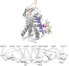
Perhaps the most studied example of a GAG–protein interaction is the binding of antithrombin to heparin and HS (Figure ). This interaction is of great pharmacological importance because heparin is widely used clinically as an anticoagulant. Binding of antithrombin to heparin has a dual effect: first, it causes a conformational change in the protein and activation of the protease inhibiting action, resulting in a 1000-fold enhancement in the rate at which it inactivates thrombin and factor Xa. Second, the heparin chain acts as a template, enhancing the physical apposition of thrombin and antithrombin. Thus, both the protease (thrombin) and the inhibitor have GAG-binding sites. Heparin acts as a catalyst in these reactions by enhancing the rate of the reaction through apposition of substrates and conformational change. After the inactivation of thrombin or factor Xa by antithrombin occurs, the complexes lose affinity for heparin and dissociate. The heparin is then available to participate in another activation/inactivation cycle. Antithrombin is a member of the serpin family of protease inhibitors, many of which bind to heparin.
(Top) Crystal structure of the antithrombin–pentasaccharide complex (from Protein Data Bank). Three structural elements that make contact with heparin are shown, including a loop (red), and two α-helices (in blue and magenta). The pentasaccharide (more...)
Early studies using affinity fractionation schemes showed that only about one-third of the chains in a heparin preparation actually bind with high affinity to antithrombin. Comparing the sequence of the bound chains with those that did not bind failed to reveal any substantial differences in composition, consistent with the later discovery that the binding site consists of only five sugar residues (Figure ) (the average heparin chain is about 50 sugar residues). Within this pentasaccharide sequence, however, a centrally located 3-O-sulfated GlcNS6S unit plays an essential role in mediating antithrombin–heparin interaction. The binding sites in GAG chains in general represent a very small segment of the chains.
Crystals of antithrombin were prepared and analyzed by X-ray diffraction to 2.6-Å resolution. The docking site for the heparin pentasaccharide is formed by the apposition of two helices, which both contain critical arginine and lysine residues at the interface (Figure ). The pentasaccharide is sufficient to activate antithrombin binding toward factor Xa, but it will not facilitate the inactivation of thrombin. For this to occur, a larger oligosaccharide of at least 18 residues is needed. As mentioned above, thrombin also contains a heparin-binding site, and the larger heparin oligosaccharide is thought to act as a template for the formation of a ternary complex with thrombin and antithrombin. In contrast to antithrombin, thrombin shows little oligosaccharide specificity. As might be expected, adding high concentrations of heparin actually inhibits the reaction, because the formation of binary complexes of heparin and thrombin or heparin and antithrombin predominate. This important principle of “activation at low concentrations and inhibition at high concentrations” also occurs in other systems in which ternary complexes form.
FGF–HEPARIN INTERACTIONS ENHANCE STIMULATION OF FGF RECEPTOR SIGNAL TRANSDUCTION
A large number of growth factors can be purified based on their affinity for heparin. The heparin-binding family of FGFs has grown to more than 22 members and includes the prototype FGF2, otherwise known as basic fibroblast growth factor. FGF2 has a very high affinity for heparin (Kd ∼ 10−9
m) and requires 1.5–2 m NaCl to elute from heparin-Sepharose. FGF2 has potent mitogenic activity in cells that express one of the FGF-signaling receptors (FGFRs; four FGFR genes are known and multiple splice variants exist). Cell-surface HS binds to both FGF2 and FGFR, facilitating the formation of a ternary complex. Both binding and the mitogenic response are greatly stimulated by heparin or HS, which promotes dimerization of the ligand–receptor complex.
The costimulatory role of HS (and heparin) in this system is reminiscent of the heparin/antithrombin/thrombin story. Indeed, the minimal binding sequence for FGF2 also consists of a pentasaccharide. However, this pentasaccharide is not sufficient to trigger a biological response (mitogenesis). For this to occur, a longer oligosaccharide (10-mer) containing the minimal sequence and additional 6-O-sulfo groups are needed to bind FGFR. The sequence that binds to both FGF2 and FGFR is prevalent in heparin but rare in HS. The requirement for this rare binding sequence reduces the probability of finding this particular arrangement in naturally occurring HS chains. Thus, some preparations of HS are inactive in mitogenesis, and those containing only one-half of the bipartite binding sequence are actually inhibitory.
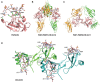
The structure of FGF2 co-crystallized with a heparin hexasaccharide has since been obtained (Figure ). The heparin fragment ([GlcNS6Sα1-4IdoA2Sα1-4]3) was helical and bound to a turn-rich heparin-binding site on the surface of FGF2. Only one N-sulfo group and the 2-O-sulfo group from the adjacent iduronic acid are bound to the growth factor in the turn-rich binding domain, and the next GlcNS residue is bound to a second site, consistent with the minimal binding sequence determined with oligosaccharide fragments. Unlike antithrombin–heparin interaction, no significant conformational change in FGF2 occurs on heparin binding. The crystal structure of acidic FGF (FGF1) has also been solved and shows similar sequences on its surface. However, the oligosaccharide sequence that binds with high affinity to FGF1 contains 6-O-sulfo groups.
Crystal and solution structures of GAG–protein complexes. (A) Crystal structure of the FGF2–hexasaccharide complex (PDB: 1BFC). Key interactions between the hexasaccharide and FGF2 are shown in black dotted lines. The hexasaccharide is (more...)
The cocrystal structure of the complex of (FGF2-FGFR)2, first solved in the absence of heparin/HS ligand, showed a canyon of positively charged amino acid residues, suggestive of an unoccupied heparin-binding site. Subsequently, the heparin–oligosaccharide-containing complex was solved after introduction of heparin oligosaccharides, suggesting a 2:2:2 complex of FGF2:FGFR1:HS (Figure ). Another important feature of this complex is the orientation of the nonreducing ends of the HS chains that terminate in an N-sulfoglucosamine residue, which arises by endolytic cleavage of chains by the enzyme heparanase (Chapter 17). The structure of the FGF1–FGFR2–HS complex is not without controversy; structural analysis of complexes formed in solution and purified by gel filtration has suggested a very different structure consisting of a 2:2:1 complex (Figure ).
CCL5–CHONDROITIN SULFATE INTERACTIONS: STABILIZATION OF A CHEMOTACTIC GRADIENT
Chemokines are another class of proteins exhibiting strong interactions with GAGs. However, these interactions do not directly affect activities of inhibitors or signaling molecules as in previous examples. Instead, they stabilize chemotactic gradients of chemokines; these gradients, in turn, direct leukocytes to sites of injury or infection. They may also facilitate cell migration through the endothelial layer of the blood vessels in which they circulate. This is no easy task; blood is flowing rapidly in these vessels and would easily destroy gradients in the absence of these interactions. CCL5, also called RANTES, is a member of a chemokine subclass that has two adjacent cysteines (CC) near its amino terminus. It is among the earliest discovered and among the most studied of the chemokines. CCL5 binds both HS and CS; but given the abundance of CS chains in normal plasma and the association of CS proteoglycans with vascular development and disease, a specific discussion of CCL5–CS interactions seems appropriate.
Although CCL5 is a small protein (∼8 kDa), it exists primarily as dimers (Kd<1 µM) that assemble into larger filamentous structures, especially in the presence of GAGs. These characteristics have made it difficult to study CCL5 structurally. Most studies have involved a mutated form (E66S) and/or study at lower pH—conditions that limit oligomerization to a dimer. Nevertheless, complementary solution data, including nuclear magnetic resonance (NMR) and small angle X-ray scattering (SAXS) have allowed construction of filament models, both with and without GAGs present. Figure shows a model of a tetramer built on the known dimer structure (PDB id, 1U4L) and complexed with CS hexamers 4-O-sulfated on all three N-acetylgalactosamine residues of the GlcAβ1-3GalNAc repeats. Individual dimers are shown as green and sea green ribbons. Additional dimers can be added to each end to build a filament structure.
The interactions between protein residues and charged groups on the CS involve a classic BBXB motif (R44, K45, N46, R47) plus a more remote arginine (R17). Each positively charged arginine is positioned to have strong electrostatic and hydrogen bond interactions with the negatively charged carboxylate of a glucuronic acid residue and 4-O-sulfo group of the following N-acetylgalactosamine residue. The positioning of these arginine residues may be unique to CS, and other GAG segments may bind quite differently. The model of the filament in Figure is also not unique, and both positioning of dimeric subunits and flexibility in CS binding geometry may allow longer GAG chains to cross-link adjacent dimers and facilitate oligomerization. A final point of note is that the groups responsible for interaction with the CCR5 receptor on leukocytes are near the amino terminus, a segment deeply buried in the dimer interface. These groups may be transiently exposed on filament dissociation to release monomers or they may be more permanently exposed on filament ends. Much additional structural work will be required to fully understand the function of CCL5.
OTHER ATTRIBUTES OF GAG–PROTEIN INTERACTIONS
In some cases, the interaction of GAG chains with proteins may depend on metal cofactors. For example, L- and P-selectins have been shown to bind to a subfraction of HS chains and heparin in a divalent-cation-dependent manner. This observation raises the possibility that other examples of cation-dependent interactions with GAG chains may exist. Indeed, annexin A2 and annexin V have been shown to bind HS in a calcium-dependent manner. The co-crystal structures of annexin A2 and annexin V with heparin oligosaccharides revealed that calcium ions can either directly or indirectly (through sequestered water) participate in the interactions with HS. Another example is calcium-dependent interaction between cytokine S100A12 and HS. In this case, formation of the functional HS-binding site requires a calcium-induced conformational change of S100A12.
Although the vast majority of GAG–protein interactions were identified and studied under neutral pH, quite a few GAG-binding proteins have been shown to interact with GAG only under acidic pH. Such proteins include histidine-rich glycoproteins, cystatin-C, selenoprotein P, and serum amyloid A. There is good reason to believe that the interactions between these proteins and GAG are physiologically relevant because extracellular environments with acidic pH exist under many physiological and pathological conditions. In fact, for these proteins, pH change essentially functions as an on/off switch of their interactions with GAG, which might bear physiological significances. Not surprisingly, histidines are often found in the GAG-binding site of these proteins because of its pH-dependent protonation of the imidazole ring.
A main technical challenge to understand GAG–protein interactions is to dissect the essential structural elements of GAGs that contribute to the binding. Unlike researchers studying DNA-binding proteins who have access to all possible DNA sequences, researchers studying GAG-binding proteins traditionally do not have access to GAG oligosaccharides with defined structures. Fortunately, this situation has begun to change in the last 5 years. With the rapid progress of chemoenzymatic and chemical synthesis (or a combination of both) of HS/CS oligosaccharides, a growing number of structural defined oligosaccharides (dp4-dp20) have become available. Many of the oligosaccharides have been used to generate microarrays for rapid determination of structural preference of GAG-binding proteins. Although we have not yet reached a stage at which we can freely design any GAG structures that we wish, the current technology can already provide a surprisingly large array of structures for structure–function studies of GAG-binding proteins.
With more than 500 GAG-binding proteins already identified (and still counting), we cannot help contemplating how this huge GAG interactome really functions at the system level. It is obvious that in any cellular environment, if one GAG-binding protein is found, it is likely that many more GAG-binding proteins are present as well. Do these GAG-binding proteins live in harmony, or are they in constant conflict by competing for binding to GAGs? When one GAG-binding protein is down-regulated, how would other GAG-binding proteins respond to this sudden availability of the free GAG-binding sites? Addressing these questions requires a systems biology approach, which has become possible with the recent increase in information about individual GAG–protein interactions. Given that spatiotemporal changes of GAG structure have profound impacts on the physiological processes associated with GAG-binding proteins, it would be natural to study this question using a systems biology approach.
ACKNOWLEDGMENTS
The authors appreciate helpful comments and suggestions from Ryan Porell, Ulf Lindahl, and So Young Kim.
FURTHER READING
Li W, Johnson DJ, Esmon CT, Huntington JA. 2004. Structure of the antithrombin–thrombin–heparin ternary complex reveals the antithrombotic mechanism of heparin. Nat Struct Mol Biol
11:
857–862. doi:10.1038/nsmb811 [
PubMed: 15311269] [
CrossRef]
Mohammadi M, Olsen SK, Goetz R. 2005. A protein canyon in the FGF–FGF receptor dimer selects from an a la carte menu of heparan sulfate motifs. Curr Opin Struct Biol
15:
506–516. doi:10.1016/j.sbi.2005.09.002 [
PubMed: 16154740] [
CrossRef]
Deshauer C, Morgan AM, Ryan EO, Handel TM, Prestegard JH, Wang X. 2015. Interactions of the chemokine CCL5/RANTES with medium-sized chondroitin sulfate ligands. Structure
23:
1066–1077. doi:10.1016/j.str.2015.03.024 [
PMC free article: PMC4456249] [
PubMed: 25982530] [
CrossRef]
Mizumoto S, Yamada S, Sugahara K. 2015. Molecular interactions between chondroitin-dermatan sulfate and growth factors/receptors/matrix proteins. Curr Opin Struct Biol
34:
35–42. doi:10.1016/j.sbi.2015.06.004 [
PubMed: 26164146] [
CrossRef]
Pomin VH, Mulloy B. 2015. Current structural biology of the heparin interactome. Curr Opin Struct Biol
34:
17–25. doi:10.1016/j.sbi.2015.05.007 [
PubMed: 26038285] [
CrossRef]
Xu D, Arnold K, Liu J. 2018. Using structurally defined oligosaccharides to understand the interactions between proteins and heparan sulfate. Curr Opin Struct Biol
50:
155–161. doi:10.1016/j.sbi.2018.04.003 [
PMC free article: PMC6078804] [
PubMed: 29684759] [
CrossRef]
Chen YC, Chen SP, Li JY, Chen PC, Lee YZ, Li KM, Zarivach R, Sun Y-J, Sue SC. 2020. Integrative model to coordinate the oligomerization and aggregation mechanisms of CCL5. J Mol Biol
432:
1143–1157. doi:10.1016/j.jmb.2019.12.049 [
PubMed: 31931012] [
CrossRef]