

NCBI Bookshelf. A service of the National Library of Medicine, National Institutes of Health.
Probe Reports from the NIH Molecular Libraries Program [Internet]. Bethesda (MD): National Center for Biotechnology Information (US); 2010-.

Chemical Genetic Analysis of Platelet Granule Secretion-Probe 2
Lynn VerPlank, Chris Dockendorff, Joseph Negri, Jose R Perez, James Dilks, Lawrence MacPherson, Michelle Palmer, Robert Flaumenhaft, and Stuart L Schreiber.
Author Information and Affiliations
Received: March 25, 2010; Last Update: February 10, 2011.
Platelet activation occurs in response to vascular injury, which triggers an array of cascading signaling events that potentiate the formation of thrombi to control bleeding. One downstream effect of activation is the secretion of granules, which plays a role in the formation of thrombi. Platelet activation inhibitors that target various surface receptors and cytosolic pathways within platelets are well validated for inhibiting thrombosis, clinically preventing adverse cardiovascular events such as intermittent claudication and stroke, and reducing mortality and morbidity in acute myocardial infarction. These inhibitors have provided a greater understanding of the mechanisms of platelet biology. Some inhibitors are currently in use in the clinical setting or in development for the treatment of thrombotic conditions. Unfortunately, many of the characterized platelet inhibitors can cause undesired side effects in patients. None of the characterized inhibitors are known to target the granule secretion machinery. We report the outcome of a high-throughput chemical library screen and a series of secondary conformation assays to identify novel, small-molecule inhibitors of platelet activation, specifically fitting into three possible attribute classes: 1) inhibitors of the pathways required for dense granule secretion, 2) inhibitors of pathways required for granule secretion, and 3) inhibitors of upstream g-coupled protein receptor (GPCR) signaling. A primary assay was developed to measure the release of granules from platelets activated through the thrombin GPCR PAR1. Of 302,457-screened compounds, 28 prioritized compounds were selected for additional characterization in secondary specificity assays, which yielded three probe candidates. These three probe candidates met the criteria as possible modulators of GPCR signaling in platelet activation. Of these, a cyanopyridone scaffold (ML160) showed acceptable potency (av. 3.75 μM ± 4.53) and inhibited P-selectin expression in a concentration-dependent manner (EC50<3 μM), as well as inhibiting SFLLRN-induced thrombus formation. This new GPCR-specific probe will be useful in future cell-based investigations of the complex mechanisms of platelet activation and in vivo studies as an antithrombotic agent in murine models.
Assigned Assay Grant No: 1 RO3 MH084076-01
Screening Center Name and PI: Broad Institute Probe Development Center, Stuart Schreiber, PhD
Chemistry Center Name and PI: Broad Institute Probe Development Center, Stuart Schreiber, PhD
Assay Submitter and Institution: Robert Flaumenhaft, Beth Israel Deaconess Medical Center, Boston, MA
PubChem Summary Bioassay Identifier (AID): AID 1678
Probe Structure and Characteristics
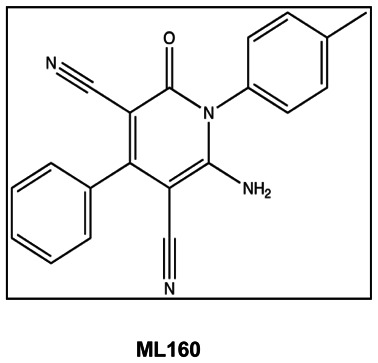
| Chemical Name | 2-amino-1-(4-methylphenyl)-6-oxo-4-phenylpyridine-3,5-dicarbonitrile |
| PubChem CID | 824820 |
| Molecular Weight | 326.35136 |
| Molecular Formula | C20H14N4O |
| AlogP | 2.9 |
| H-Bond Donor | 1 |
| H-Bond Acceptor | 4 |
| Rotatable Bond Count | 2 |
| Exact Mass | 326.116761 |
| Topological Polar Surface Area | 93.9 |
| CID/ML No. | Target Name | IC50/EC50 (nM) [SID, AID] | Anti-target Name(s) | IC50/EC50 (μM) [SID, AID] | Fold Selective* | Secondary Assay(s) Name: IC50/EC50 (nM) [SID, AID] |
|---|---|---|---|---|---|---|
| 824820/ML160 | Granule secretion | 361 [SID 87556782, AID 2398, AID 2519] | Luciferase | 112 [SID 1738663, AID 1891] | 310 | P-selectin 3000 [SID 87219012, AID 2518, AID 2511]; cAMP [SID 17386663, AID 2645, AID 2646]; FITC Phalloidin [SID 17386663, AID 2655, AID 2657]; PMA [SID 17386663, AID 2529, AID 2509]; Ca Ionophore [SID 17386663, AID 2522, AID 2527] |
- *
Selectivity = anti-target IC50/target IC50.
Recommendations for Scientific Use of the Probe
The goal of this project is to identify novel probes that inhibit platelet activation. This project focuses on the pathways involved in the activation of platelets initiated through the thrombin receptor pathway. The SFLLRN peptide activates platelets through the G-protein coupled receptor (GPCR) protease-activated receptor-1 (PAR1). The SFLLRN peptide is derived from the cleavage of the N-terminus of the PAR1 receptor by thrombin (1). Upon activation, the platelets undergo many changes induced by multiple signaling cascades. One downstream effect of activation is the secretion of granules, which then potentiate platelets in controlling bleeding. Granule secretion also contributes to the growth of thrombi, which if not controlled properly can cause blockage of blood vessels and trigger myocardial infarction (MI). Probes from this project are defined as fitting into three possible attribute classes: 1) inhibitors of the pathways required for dense granule secretion, 2) inhibitors of the pathways required for granule secretion, which includes dense granules, alpha-granules, and lysosomes, and 3) inhibitors of upstream GPCR signaling. The first and second classes of inhibitors target distal mechanisms of secretion (i.e., the platelet second messenger systems) (2). The third class of inhibitors targets the proximal cell-surface receptors.
The probes identified in this project will be used to further investigate the complex mechanisms of platelet activation. As these probes have been classified as GPCR-specific, additional assays developed in the Assay Provider’s laboratory (Robert Flaumenhaft, MD) will be performed to determine the specific GPCR target and site of action on the target GPCR signaling pathway. Depending on the results of these additional assays, these probes could be evaluated for their potential as antithrombotic agents in an in vivo murine model.
The GPCR-specific probe presented in this report is a valuable tool compound with unique attributes. Additional experimentation carried out after identification of the probe has demonstrated that this compound acts specifically on the GPCR PAR1. It exhibits several characteristics that distinguishes it from published orthosteric inhibitors of PAR1. This area of investigation is of particular interest to the Assay Provider and others studying clinical aspects of platelet biology as platelets represent a good cellular target for pharmacological manipulation via allosteric modulation (3). The central challenge in developing an antiplatelet agent is how to achieve a potent antiplatelet effect while avoiding hemorrhagic complications. Development of modulators of platelet function with novel modes of action, such as the probe described here, could limit bleeding complications associated with current antiplatelet agents (4).
1. Introduction
Platelets are small, anucleate cells that circulate in the blood of mammals, and are required for maintaining hemostasis. The number of platelets circulating and their relative activity requires strict control. Too few platelets or too little activity can cause uncontrolled bleeding, while too many platelets and too much activity can cause acute thrombosis formation. Thrombi formation can block blood vessels causing myocardial and cerebral infarctions. Platelets undergo dramatic phenotypic changes in response to vascular injury that lead to the formation of thrombosis. These phenotypic changes are the result of an activation process induced by a diverse array of agonists (e.g., thrombin, adenosine diphosphate [ADP], epinephrine, and thromboxane A2) that act on platelet surface receptors. Phenotypic changes include granule secretion, shape change, and expression of specific cell-surface proteins. Platelet activation inhibitors are well validated for the inhibition of thrombosis and the clinical prevention of adverse cardiovascular events, including MI and stroke (5). These inhibitors target various platelet G-protein coupled receptors (GPCRs), as well as certain cytosolic enzymes, which comprise key parts of the platelet activation pathway. Examples of important drugs within the category of GPCR inhibitors include: clopidogrel (Plavix®, Bristol-Myers Squibb/Sanofi Aventis), the metabolism of which leads to irreversible inhibition of the ADP receptor P2Y12; and SCH 530348, a potent inhibitor of protease activated receptor (PAR1) currently in Phase 3 clinical trials. Examples of platelet enzyme inhibitors include: aspirin, an irreversible inhibitor of COX1; and cilostazol (Pletal®, Otsuka), a cyclic nucleotide phosphodiesterase 3 (PDE3) inhibitor in clinical use for intermittent claudication. The structures of these compounds are depicted in Figure 1.

Figure 1
Examples of Platelet Activation Inhibitors.
Unfortunately, many of these drugs and others within the broader category of antithrombotics suffer from significant liabilities. For example, P2Y12 inhibition leads to a significant risk of dangerous hemorrhagic side effects (6). Key platelet enzymes such as COX1 and PDE3 are also present in many other cell types, and their inhibition can lead to undesired complications (7). For this reason, novel inhibitors of platelet activation are of great interest, particularly those that modulate new targets, those that inhibit select features of platelet activation, and those that demonstrate novel pharmacological properties such as allosteric inhibition (3).
Platelets are best studied via chemical genetic analysis due to their anucleate state rendering them unsusceptible to direct genetic manipulation. Platelets in plasma are a readily available reagent in the form of expired donor-collected units of platelet-rich plasma (PRP) from blood centers nationwide. The Assay Provider’s laboratory previously developed and executed a small molecule screen to study platelet activation in PRP using granule secretion as a measurement of platelet activation (2). The assay measures the amount of ATP released into the plasma via secreted dense granules upon activation (8).
The purpose of these studies is to use a cell-based platelet assay to identify novel inhibitors of platelet activation. Inhibitors identified from this project are defined as fitting into three possible attribute classes: 1) inhibitors of the pathways required for dense granule secretion, 2) inhibitors of the pathways required for granule secretion, which includes dense granules, alpha-granules, and lysosomes, and 3) inhibitors of upstream GPCR signaling. The first and second classes of inhibitors target distal mechanisms of secretion (i.e., the platelet second messenger systems) (2). There are no known compounds that selectively inhibit granule secretion in either the first or second class. The third class of inhibitors targets the proximal cell-surface receptors. One important platelet GPCR mentioned above is PAR1, which undergoes a cleavage reaction by the serine protease thrombin that allows its tethered peptide ligand to agonize the receptor (9).
The primary screen was adapted from the cell-based phenotypic screen previously executed by the Assay Provider’s laboratory with an aim to identify inhibitors of platelet activation (2). Platelets were activated with the thrombin receptor activation peptide SFLLRN, which binds the PAR1 GPCR (1). Upon activation, the platelets undergo many changes induced by multiple signaling cascades, which results in the secretion of granules. Platelet activation was quantified by measurement of ATP from the secreted granules, as measured by a luciferin-luciferase assay. Cilostazol, a known platelet aggregation inhibitor that targets PDE3, was used as a positive control in the primary assay (see Figure 1). It was chosen as a positive control for the high level of inhibition seen in the primary assay, although it acts on a target that is not relevant to this project.
The secondary assays in this project served to determine the specificity of the inhibitors identified in the primary assay. Specifically, the first secondary assay distinguishes between inhibitors of granule secretion and those acting nonspecifically on the ATP-detection system. From a list of compounds acting specifically to decrease the levels of secreted ATP-rich granules from platelets, those compounds known to inhibit intracellular platelet targets were filtered out. The final secondary assays were designed to differentiate between inhibitors of the distal granule secretion events and those inhibiting proximal GPCRs.
2. Materials and Methods
Materials and Reagents
Expired units of platelets were acquired from BloodSource (Sacramento, CA), Rhode Island Blood Center (Providence, RI), Blood Center of New Jersey (East Orange, NJ), and Blood Center of Wisconsin (Milwaukee, WI). CellTiter-Glo was purchased from Promega (Madison, WI). SFLLRN peptide was purchased from Bachem (Torrance, CA), PBS was acquired from Broad Institute Supply and Quality Management Group (SQM), and Cilostazol, ATP, and Resveratrol were acquired from Sigma. White, solid-bottom, flat, opaque 384-well plates were purchased from Aurora Biotechnologies (Catalog no. 00030721).
2.1. Assays
A summary listing of completed assays and corresponding PubChem AID numbers is provided in Table 1 (see Appendix 1).
2.1.1. Primary Screen and Conformation at Dose: Granule Secretion
Refer to Appendix 2 for the detailed assay protocol.
Platelet volumes from a single donor ranged from 200 mL to 700 mL. Samples were taken for counting platelets/mL and tested for activation activity by the addition of SFLLRN and CellTiter-Glo. Only platelets demonstrating a high level of activation were used in the assay. The 384-well plates were filled with 20 μL/well with platelets, using a Thermo Multidrop Combi liquid handler in a biosafety cabinet. The volume of platelets determined the number plates used for a run. The filled plates were loaded into an automated incubator set at 30°C, 95% humidity, 5% CO2, docked to an enclosed automated screening system (HighRes Biosolutions, Woburn, MA). Unsealed compound plates were loaded into another automated incubator on the same system, set at 22°C, 15% humidity. CellTiter-Glo substrate was resuspended in PBS (100 mL/bottle, 1:1) and the SFLLRN peptide was reconstituted in PBS to 10 mM. The CellTiter-Glo/SFLLRN screening solution was prepared by dilution of CellTiter-Glo solution to 1:4 with PBS and the addition of reconstituted SFLLRN to a final concentration of 15 μM. Each run was initiated by the software application Broad Chemical Biology Informatics Platform (CBIP) and scheduled with Cellario (HighRes Biosolutions, Woburn, MA). Robotic arms moved plates from the different instruments on the screening system. Compounds were pinned from DMSO daughter plates into assay plates using a MicroPin (HighRes Biosolutions, Woburn, MA). After compound addition, assay plates were returned to the incubator for a 30-minute incubation. At the completion of incubation, plates were moved to a MultiDrop Combi for addition of 10 μL CellTiter-Glo/SFLLRN solution per well, to a final well concentration of 0.083X CellTiter-Glo and 5 μM SFLLRN. The plates were incubated on the deck of the screening system for 15 minutes, at room temperature. At the completion of incubation, the plates were read for luminescence in an EnVision 2104 Multilabel Reader (Perkin Elmer, Waltham, MA). The ultra sensitive detection was used, with the 1536 aperture in place, to decrease bleed-through from adjacent wells. Read time was 0.1s/well. The same assay protocol was used for a conformation at dose response. The 1584 cherry-pick compounds were received from BioFocus and powder compounds tested in this assay were either synthesized (BIPDeC) or purchased from various vendors.
2.1.2. Luciferase Counterscreen
Refer to Appendix 2 for the detailed assay protocol.
ATP was diluted to 1.5 μM in PBS from 10-mM aliquots in PBS. Next, 1:1 aliquots of CellTiter-Glo (Promega), reconstituted in PBS, were diluted to 1:4 with PBS. A MultiDrop Combi (Thermo) was used to add 20 μL of 1.5 μM ATP in PBS. Then, 50 nL of compounds at dose were added to the assay plates using a CyBi-Well (CyBio Inc.). Upon completion of compound addition, 10 μL of CellTiter-Glo (1:4) in PBS was added to every well, to a final concentration of 0.083X. Plates were transferred to an EnVison Plate Reader (PerkinElmer, Waltham, MA) to measure luminescence. The ultra sensitive detection was used, with the 1536 aperture in place, to decrease bleed-through from adjacent wells. Read time was 0.1s/well.
2.1.3. Secondary Assay for 14C -Serotonin Release
This assay was performed in the Assay Provider’s laboratory with 28 cherry-pick compounds acquired from BioFocus, selected from a narrowed list of putative platelet activation inhibitory compounds. Platelet rich plasma (PRP; 4.5 mL, approximately 3×10⁸ platelets/mL) was incubated in the presence of 14C-serotonin (0.225 μCi) for 45 minutes at 37°C. Next, 15% acid-citrate-dextrose (ACD)+ 150 nM prostaglandin E1 (PGE1) was added to the PRP and spun at 1000 g for 10 minutes to maintain the resting state of platelets while removing unincorporated 14C-serotonin. The platelets were resuspended in HEPES-Tyrodes buffer (10 mM HEPES/NaOH, pH 7.4, 5.56 mM glucose, 137 mM NaCl, 12 mM NaHCO3, 2.7 mM KCl, 0.36 mM NaH2PO4, 1 mM MgCl2) to a platelet concentration of 2×10⁸ platelets/mL. Compounds were added to resuspended platelets at 10 μM and incubated for 15 minutes. Platelets were subsequently stimulated with 5 μM SFLLRN peptide and 5 μM imipramine (to prevent 14C-serotonin reuptake) for 15 minutes. The platelets were separated from medium by centrifugation at 8000 × g. Supernatants were collected and counted using a beta liquid scintillation counter. Disintegrations per minute (DPM) were collected for 50 μL of platelets.
2.1.4. Secondary Assay for SFLLRN-induced P-selectin Surface Expression
This assay was also performed in the Assay Provider’s laboratory with 28 cherry-pick compounds acquired from BioFocus, selected from a narrowed list of putative platelet activation inhibitory compounds. In addition to the 28 cherry-pick compounds acquired from BioFocus, powder compounds synthesized (BIPDeC) or purchased from various sources were screened in this and the following assays in the Assay Provider’s laboratory. Washed platelets obtained from individual donors were treated with a selection of compounds resourced as powders found to be active in secondary assays. Platelet samples were tested at a range of concentrations from 0 μM to 100 μM of compounds. Following compound addition, platelets were subsequently stimulated with 5 mM SFLLRN. After a 15-minute incubation, phycoerythrin-labeled anti-P-selectin antibody (BD Biosciences) was added for a 20-minute incubation. The samples were analyzed by flow cytometry to determine P-selectin expression on the surface of the platelets as a response to activation. Geometric mean values were collected for each sample.
2.1.5. Counterscreen for Identification of Inducers of cAMP in Platelets
Washed platelets obtained from individual donors were treated with a selection of compounds that were chosen based on activity in an SFLLRN-induced P-selectin expression assay. Prostaglandin E1 (PGE1) at 1 μM was used as a positive control to elevate platelet cAMP levels. Compounds were tested at 10 μM. Platelets were lysed following compound or control incubation, and lysates were analyzed with a cAMP competitive ELISA kit (Thermo Scientific). Values were converted to cAMP concentration (pmol/mL) based on a cAMP standard curve measured in parallel.
2.1.6. Secondary Assay for Identification of Inhibitors SFLLRN-mediated Actin Polymerization in Platelets
Platelets were obtained from individual donors, gel-filtered, and treated with DMSO (neutral control) or compounds (10 μM) that did not induce elevation of cAMP in platelets as measured in a cAMP assay. Following incubation with compounds, platelets were either treated or not treated with the thrombin receptor peptide agonist, SFLLRN. Both populations of platelets were fixed, and then incubated in the presence of FITC-phalloidin and Triton X-100. Samples were analyzed by flow cytometry to determine levels of actin polymerization upon activation. Actin filament formation results in higher fluorescent measurements. Geometric mean values were collected for each sample.
2.1.7. Counter Assay for Identification of Inhibitors of Ca2+-ionophore (A23187)-induced P-selectin Surface Expression
Washed platelets obtained from individual donors were treated with compounds found to show less than 50% inhibition in an SFLLRN-induced FITC phalloidin assay. Platelet samples were incubated with 30 μM of compound. Following compound addition, platelets were stimulated with 10 μM A23187. After a 15-minute incubation, phycoerythrin-labeled anti-P-selectin antibody (BD Biosciences) was added for a 20-minute incubation. The samples were analyzed by flow cytometry to determine P-selectin expression on the surface of the platelets as a response to activation. Geometric mean values were collected for each sample.
2.1.8. Counter Assay for Identification of Inhibitors of Phorbol Myristate Acetate (PMA)-induced P-selectin Surface Expression
Washed platelets obtained from individual donors were treated with compounds found to show less than 50% inhibition in an SFLLRN-induced FITC phalloidin assay. Platelet samples were treated with 30 μM of compound. Following compound addition, platelets were stimulated with 5 nM PMA. After a 15-minute incubation, phycoerythrin-labeled anti-P-selectin antibody (BD Biosciences) was added for a 20-minute incubation. The samples were analyzed by flow cytometry to determine P-selectin expression on the surface of the platelets as a response to activation. Geometric mean values were collected for each sample.
2.2. Probe Chemical Characterization
After preparation as described in Section 2.3, the probe (ML160, CID 824820, SID 87556782, MLS002699917) was analyzed by HPLC, both 1H and 13C NMR, and high-resolution mass spectrometry. The data were consistent with the structure of the probe, and HPLC indicated an isolated purity of greater than 99%. The relevant data is provided below. The observed solubility of the probe in PBS buffer (pH 7.4) was determined to be 1.6 μM. Aqueous stability in PBS was monitored over a 48-hour period, and the data is provided in Figure 2, indicating excellent aqueous stability. Plasma protein binding (PPB) was determined to be 96% bound in human plasma and 91% bound in murine plasma. The probe is stable in human and murine plasma with essentially 100% remaining after a 5-hour incubation period. The solubility, PPB, and plasma stability results are summarized in Table 2 (see Section 3.4). The probe and five additional analogs were submitted to the SMR collection [MLS002699917 (probe), MLS002699918, MLS002699919, MLS002699920, MLS002699921, and MLS002699923 (analogs)].

Figure 2
Probe Stability in PBS at 23°C.
2.3. Probe Preparation
Chemistry Experimental Methods
The probe and the analogs of the cyanopyridone class were made via an efficient 2-pot process, without requiring chromatography in any step. 2-cyanoacetic acid was coupled with a series of primary amines, and the resulting amides were subjected to Knoevenagel condensations with several different benzaldehydes. The resulting enamides were condensed with malononitrile; stirring in the air led to the formation of the desired aromatic cyanopyridones (10,11), which readily precipitated and were washed with various solvents to give materials with excellent purity in most cases. The synthesis of the probe is provided in Scheme 1.

Scheme 1
Synthesis of Cyanopyridone Probe.
General Details. All reagents and solvents were purchased from commercial vendors and used as received. NMR spectra were recorded on a Bruker 300 MHz or Varian UNITY INOVA 500 MHz spectrometer as indicated. Proton and carbon chemical shifts are reported in parts per million (ppm;δ) relative to tetramethylsilane, CDCl3 solvent, or d6-DMSO (1H δ 0, 13C δ 77.0, or 13C δ 39.5, respectively). NMR data are reported as follows: chemical shifts, multiplicity (obs. = obscured, br = broad, s = singlet, d = doublet, t = triplet, m = multiplet); coupling constant(s) in Hz; integration. Unless otherwise indicated, NMR data were collected at 25°C. Flash chromatography was performed using 40–60 μm Silica Gel (60 Å mesh) on a Teledyne Isco Combiflash Rf system. Tandem liquid chromatography/mass spectrometry (LCMS) was performed on a Waters 2795 separations module and Waters 3100 mass detector. Analytical thin layer chromatography (TLC) was performed on EM Reagent 0.25 mm silica gel 60-F plates. Visualization was accomplished with UV light and aqueous potassium permanganate (KMnO4) stain followed by heating. High-resolution mass spectra were obtained at the MIT Mass Spectrometry Facility with a Bruker Daltonics APEXIV 4.7 Tesla Fourier Transform Ion Cyclotron Resonance mass spectrometer. Compound purity and identity were determined by UPLC-MS (Waters, Milford, MA). Purity was measured by UV absorbance at 210 nm. Identity was determined on a SQ mass spectrometer by positive electrospray ionization. Mobile Phase A consisted of either 0.1% ammonium hydroxide or 0.1% trifluoroacetic acid in water, while mobile Phase B consisted of the same additives in acetonitrile. The gradient ran from 5% to 95% mobile Phase B over 0.8 minutes at 0.45 mL/min. An Acquity BEH C18, 1.7 μm, 1.0 × 50 mm column was used with column temperature maintained at 65°C. Compounds were dissolved in DMSO at a nominal concentration of 1 mg/mL, and 0.25 μL of this solution was injected.
Step 1. Preparation of 2-cyano-N-(p-tolyl)acetamide: p-Toluidine (2.14 g, 20.0 mmol) and 2-cyanoacetic acid (1.79 g, 21.0 mmol) were sealed in a flask with a stir bar and dissolved with tetrahydrofuran (THF, 67 mL) to form a pale yellow solution. Next, 1-ethyl-3-(3-dimethylaminopropyl) carbodiimide (EDCI; 4.41 g, 23.0 mmol) was added, and the reaction was stirred for 22 h. The mixture was then poured onto water (500 mL) in an Erlenmeyer flask, stirred for 1 h, then filtered and washed with water. The white solid was dried under suction for 30 minutes (min), then dried under high vacuum to yield the title compound (3.10 g, 89%) in excellent purity (LCMS). The material was used immediately in the subsequent step without further characterization.
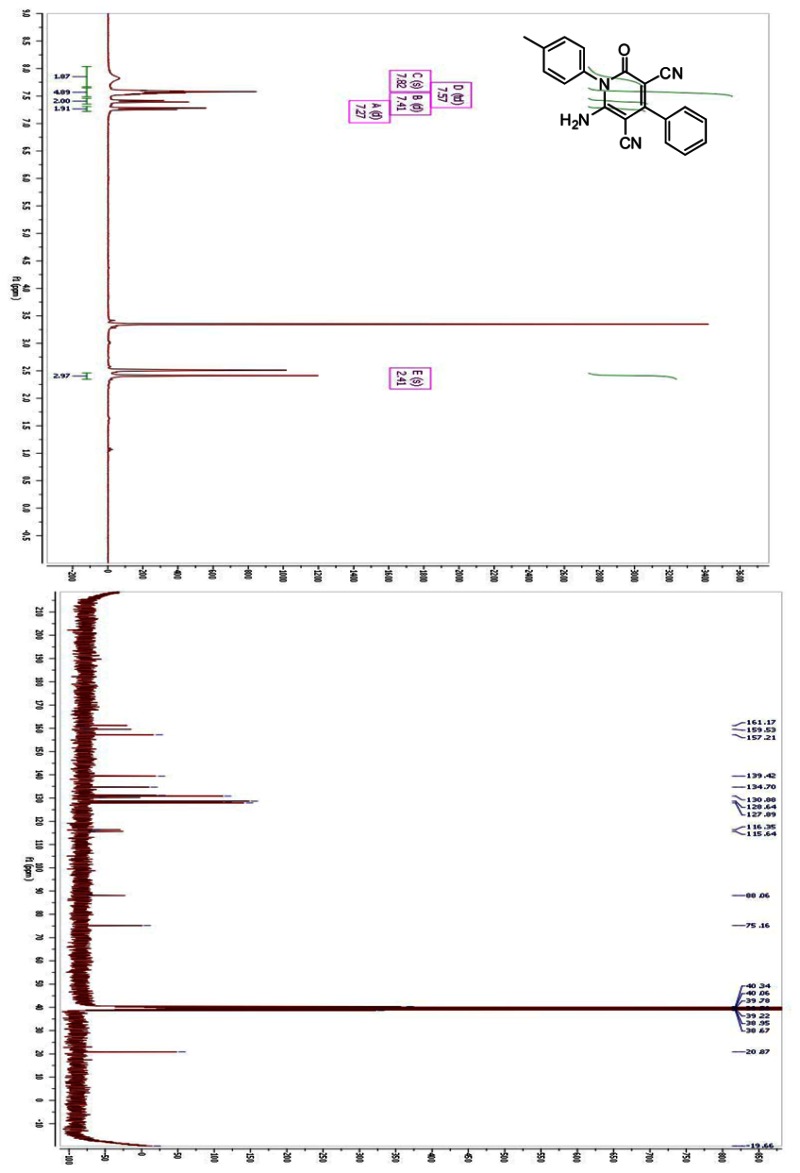
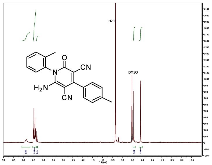
Steps 2 and 3. Preparation of the Probe (6-amino-2-oxo-4-phenyl-1-(p-tolyl)-1,2-dihydropyridine-3,5-dicarbonitrile). 2-cyano-N-p-tolylacetamide (697 mg, 4.00 mmol) and ethanol (13.3 mL) were added to flask with a stir bar, forming a white suspension. Benzaldehyde (0.554 mL, 4.20 mmol) and piperidine (four drops by Pasteur pipette) were then added. A water condenser was attached to the flask, and the reaction was refluxed for 5 h. During this time a yellow solid gradually precipitated. After 5 h, LCMS analysis indicated complete conversion to the intermediate Knoevenagel adduct. Malononitrile (277 mg, 4.20 mmol) and more piperidine (0.40 mL, 4.00 mmol) were added, and the yellow suspension was stirred under air for 17 h. The resulting white solid was filtered and washed with ice-cold ethanol, then dried under high vacuum to yield the title compound (627 mg, 48%). 1H NMR (300 MHz, d6-DMSO): δ 7.82 (br s, 1 H), 7.62–7.51 (m, 5 H), 7.40 (d, J = 8.3 Hz, 2 H), 7.26 (d, J = 8.3 Hz, 2 H), 2.41 (s, 3 H). 13C NMR (75 MHz, d6-DMSO): δ 161.2, 159.5, 157.2, 139.4, 134.7, 131.2, 130.9, 130.3, 128.6, 128.2, 127.9, 116.4, 115.6, 88.0, 75.2, 20.87. HRMS (ESI+): calculated mass for C17H17BrN2O2 [M+H] 361.0546, found 361.0532.
2.4. Additional Analytical Analysis
Plasma Protein Binding. Plasma protein binding was determined by equilibrium dialysis using the Rapid Equilibrium Dialysis (RED) device (Pierce Biotechnology, Rockford, IL) for both human and mouse plasma. Each compound was prepared in duplicate at 5 μM in plasma (0.95% acetonitrile, 0.05% DMSO) and added to one side of the membrane (200 μL) with PBS pH 7.4 added to the other side (350 μL). Compounds were incubated at 37°C for 5 h in a 250 rpm orbital shake. Following the incubation, the samples were analyzed by UPLC-MS (Waters, Milford, MA) with compounds detected by SIR detection on a single quadrupole mass spectrometer.
Plasma Stability. Plasma stability was determined at 37°C for 5 h in both human and mouse plasma. Each compound was prepared in duplicate at 5 μM in plasma diluted 50/50 (v/v) with PBS pH 7.4 (0.95% acetonitrile, 0.05% DMSO). Compounds were incubated at 37°C for 5 h with a 250 rpm orbital shake with time points taken at 0 and 5 h. Samples were analyzed by UPLC-MS (Waters, Milford, MA) with compounds detected by SIR detection on a single quadrupole mass spectrometer.
3. Results
3.1. Summary of Screening Results
A high throughput screen (HTS) of 302,457 compounds (AID 1663) was performed in duplicate to measure platelet activation by monitoring granule secretion in PRP. Using expired PRP activated with the thrombin receptor activation peptide SFLLRN, platelet activation was quantified by measurement of ATP from secreted granules, as measured by a luciferin-luciferase assay. A new assay format was developed for this project to aid in scaling up to screen the full MLPCN library with the use of automation. A stable luciferin-luciferase reagent (CellTiter-Glo, Promega) was adapted to measure secreted granules without lysing the platelets. Compounds were screened at a final concentration of 7.5 μM. From the HTS data, 661 compounds were classified as hits, based on inhibiting granule secretion ≥ 50%. A set of 1584 compounds was chosen as cherry picks, including available positive compounds, compounds classified as inconclusive, and additional inactive compounds containing structures similar to active compounds to provide additional SAR information. Cherry-picked compounds were retested in a concentration-dependent manner in the primary assay using PRP activated with SFLLRN (AID 1889). Out of 1584 compounds, 651 compounds were confirmed as active in this assay with IC50 values < 10 μM. The same set of dose-response compound plates were also tested in a biochemical counterscreen to rule out activity seen in the primary assay due to nonspecific inhibition from the luciferin-luciferase detection system (AID 1891). The 446 compounds showing an IC50 > 112.2 μM in the counterscreen were considered inactive in this assay, thus considered specific for inhibiting platelet activation. The results from these secondary assays were taken together to narrow the list to specific active compounds by advancing only compounds that exhibited a ratio of IC50 values of greater than 10-fold (i.e., counter/primary) from both assays. From this list, compounds that had been previously shown to inhibit platelet activation were excluded. Of the remaining compounds, 30 compounds were prioritized for additional characterization in secondary assays. Of these 30 prioritized compounds, 28 were acquired thorough an additional cherry pick and tested in a suite of secondary specificity assays in the Assay Provider’s laboratory (see Table 1, Appendix 1; Figure 3). These assays narrowed the list of compounds to four. The four probe candidates and analogs of each were acquired as dry powders and retested in the confirmatory and secondary assays. Of these, one of the four probe candidates did not retest and was removed from further consideration as a probe. Completion of the series of secondary assays confirmed that the three remaining compounds met the criteria for a Class III probe, possible modulators of GPCR signaling in platelet activation (see Table 1, Appendix 1; Figure 3).
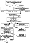
Figure 3
Critical Path for Probe Development.
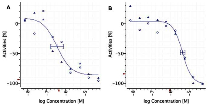
The second of the three probes to meet the defined probe criteria is a cyanopyridone (ML160, CID 824820). This probe reproducibly retests in the primary assay yielding IC50 values of 0.36 μM and 4.35 μM (see Figure 4) with improved potency over the positive control cilostazol. Variability in IC50 values was observed across experiments and was attributed to differences in platelet response from donor to donor; however, the rank ordering of potency within a probe series was maintained across multiple experiments. The sample (SID 87556782) described in Figure 4 (see Section 3.2) was tested in four different donor samples. The IC50 values from these experiments range from 0.36 μM to 9.93 μM with an average IC50 of 3.75 μM ± 4.53 (data not shown). The cyanopyridone probe also inhibits P-selectin expression in a concentration-dependent manner, with an EC50 less than 3 μM. This result demonstrated that the probe candidate met Class II or Class III probe definitions (see Figure 3). A cyclic AMP (cAMP) assay performed on the cyanopyridone probe demonstrated that the compound did not have activity in modulating cAMP levels, as compared to the potent activity of the positive control compound cilostazol (see Figure 1). Additional testing of the probe candidate in a fluorescent actin polymerization assay determined that it has greater than 50% activity in inhibiting SFLLRN-induced actin filament formation. The results of this assay suggest the probe is acting as a potential Class III probe, at an upstream GPCR target. Counterscreening in two additional assays in which platelets were activated with phorbol myristate acetate (PMA) or Ca2+ ionophore, agents that stimulate activation outside the GPCR signaling pathways, demonstrated that the cyanopyridone meets the criteria as a Class III probe (see Table 2, Section 3.4; Figure 3).
3.2. Dose Response Curves for Probe
3.3. Scaffold/Moiety Chemical Liabilities
The scaffold does not have obvious chemical liabilities. It is stable over 48 h in PBS buffer and shows excellent stability in both human and mouse plasma. As of November 2010, the probe (CID 824820) was active in seven assays out of 390 reported in PubChem, of which six assays are related to this platelet activation project. Compounds of the cyanopyridone substructure have only been reported to be active in one other screening project listed in PubChem (inhibition of mitochondrial fusion, AID 1362); therefore, the probe is considered not to be a promiscuous binder.
3.4. SAR Tables
The structures and the activities of the probe and analogs in the primary assay and the suite of secondary assays are summarized in Table 2.
Table 2SAR Analysis and Properties of Cyanopyridone Probe and Analogs
| SAR Analysis |  | Target Activity (μM) | Anti-target Activity (@ 30 μM) | ||||||||
|---|---|---|---|---|---|---|---|---|---|---|---|
| No | CID/SID | Broad ID | * | R1 | R2 | R3 | EC50 (Dense Granule Release) a | EC50 (P-Selectin expression) | % Inhib. SFLLRN-induced actin polym. @10 μM | % Inhib. PMA-induced P-selectin expression | % Inhib. Ca-ionophore-induced P-selectin expression |
| 1 | 824820/ 87556782 | BRD-K5077615 2-001-07-0 | S | 4-MePh | Ph | H | 0.4 | <3 | 99* | 6* | 38* |
| Solubility (PBS): 1.6 μM; Plasma Protein Binding (Human, Mouse): 96, 91%, Plasma Stability (Human, Mouse): 100, 100%, Purity: >99% | |||||||||||
| 2 | 1034641 87556784 | BRD-K12168891 -001-05-7 | S | 4-MePh | 4-ClPh | H | 0.6 | <3 | NT | NT | NT |
| Solubility (PBS): 11.5 μM; Plasma Protein Binding (Human, Mouse): 97, 93%, Plasma Stability (Human, Mouse): 100, 96%, Purity: 96% | |||||||||||
| 3 | 12425537 87556786 | BRD-K12238169 -001-01-2 | S | Me | Ph | H | 1.7 | NT | NT | NT | NT |
| Solubility (PBS): 24 μM; Plasma Protein Binding (Human, Mouse): 76, 63%, Plasma Stability (Human, Mouse): 100, 96%, Purity: >99% | |||||||||||
| 4 | 44629412 87556783 | BRD-K12621773 -001-01-7 | S | 2-MePh | 4-MePh | H | 2.0 | NT | NT | NT | NT |
| Solubility (PBS): 6.8 μM; Plasma Protein Binding (Human, Mouse): 97, 91%, Plasma Stability (Human, Mouse): 100, 98%, Purity: 99% | |||||||||||
| 5 | 1034643 87556785 | BRD-K29715937 -001-02-9 | S | 4-ClPh | 4-MePh | H | 9.2 | <3 | NT | NT | NT |
| Solubility (PBS): 0.5 μM; Plasma Protein Binding (Human, Mouse): 97, 92%, Plasma Stability (Human, Mouse): 100, 98%, Purity: 99% | |||||||||||
| 6 | 44629408 87556787 | BRD-K84855052 -001-01-6 | S | 4-MePh | Ph | Ac | >50 | NT | NT | NT | NT |
| Solubility (PBS): 233 μM; Plasma Protein Binding (Human, Mouse): 97, 88%, Plasma Stability (Human, Mouse): 100, 89%, Purity: >99% | |||||||||||
NT = not tested;
- *
S = synthesized; P= purchased;
- a
Primary HTS assay
The probe (entry 1) demonstrated the highest level of inhibition, but comparable activity was also observed when a para-chloride was added to the “eastern” ring (entry 2). The activity was attenuated by replacement of the N-para-tolyl group of the probe with ortho-tolyl (entry 4). The N-methyl analog (entry 3) retained respectable (but decreased) activity. Acylation of the pendant amino group of the probe greatly decreased the activity (entry 6).
3.5. Cellular Activity
The primary assay and many of the secondary assays were performed on platelets in plasma or washed platelets (see Section 2.1 for experimental details). Those compounds found to be toxic to platelets would have demonstrated no effect or an increased luminescence response in the primary assay and were not selected for additional analysis.
3.6. Profiling Assays
As described in Section 3.3, members of the cyanopyridone substructure present in the MLSMR collection have only been reported to be active in one other screening project (inhibition of mitochondrial fusion). Other profiling screens (e.g., CEREP) have not been performed with this compound.
4. Discussion
4.1. Comparison to Existing Art and How the New Probe is an Improvement
Additional studies beyond the scope of the outlined project have demonstrated this probe exhibits several characteristics that distinguish it from existing inhibitors of the platelet GPCR PAR1. It inhibits platelet secretion and aggregation without inhibiting platelet shape change (data not shown), suggesting that it inhibits Gαq, but not Gα12/13 signaling (9). Further studies in the Assay Provider’s laboratory are required to determine the specific mode of action of this probe, which are ongoing. It is possible the probe may be acting in an allosteric manner. If this were the case, this would be an improvement over the existing art. Currently, there are no known allosteric inhibitors of PAR1 with the ability to selectively inhibit certain phenotypic changes (e.g., granule secretion, but not platelet shape change). This probe could have unique advantages over orthosteric GPCR inhibitors currently used as antithrombotic drugs (3).
4.2. Mechanism of Action Studies
The Assay Provider’s laboratory is currently investigating the mechanism of action of this probe. The proposed allosteric mechanism of PAR1 inhibition will be studied by measuring the agonist dose/response with different probe concentrations. Certain shifts in the agonist dose/response curves will support an allosteric, noncompetitive mechanism of inhibition. Mutational studies to determine the GPCR binding site(s) are also underway.
4.3. Planned Future Studies
Concurrent with the mechanistic studies described above, the Broad Institute is planning to synthesize and further optimize analogs in a different probe series, based on the 1,3-diaminophenyl (DAP) scaffold described in Chemical Genetic Analysis of Platelet Granule Secretion-Probe 3 report. This strategy is based on ongoing studies of thrombin-induced platelet aggregation (not presented in this report). These analogs will be tested in assays designed and executed in the Assay Provider’s laboratory, with the goal of finding analogs with enhanced potencies. Promising compounds will also be tested in mouse models of thrombus formation.
5. References
- 1.
- Vu TK, Hung DT, Wheaton VI, Coughlin SR. Molecular cloning of a functional thrombin receptor reveals a novel proteolytic mechanism of receptor activation. Cell. 1991 Mar 22;64(6):1057–68. [PubMed: 1672265]
- 2.
- Flaumenhaft R, Sim DS. The platelet as a model for chemical genetics. Chem Biol. 2003 Jun;10(6):481–6. [PubMed: 12837380]
- 3.
- Dowal L, Flaumenhaft R. Targeting platelet G-protein coupled receptors (GPCRs): looking beyond conventional GPCR antagonism. Curr Vasc Pharmacol. 2010 Mar;8(2):140–54. [PubMed: 19485898]
- 4.
- Shehab N, Sperling LS, Kegler SR, Budnitz DS. National estimates of emergency department visits for hemorrhage-related adverse events from clopidogrel plus aspirin and from warfarin. Arch Intern Med. 2010 Nov 22;170(21):1926–33. [PubMed: 21098354]
- 5.
- Michelson AD. Antiplatelet therapies for the treatment of cardiovascular disease. Nat Rev Drug Discov. 2010 Feb;9(2):154–69. [PubMed: 20118963]
- 6.
- Wiviott SD, Braunwald E, McCabe CH, Montalescot G, Ruzyllo W, Gottlieb S, Neumann FJ, Ardissino D, De Servi S, Murphy SA, et al. for the TRITON-TIMI 38 Investigators. Prasugrel versus clopidogrel in patients with acute coronary syndromes. N Engl J Med. 2007 Nov 15;357(20):2001–15. Epub 2007 Nov 4. [PubMed: 17982182]
- 7.
- Ding B, Abe J, Wei H, Huang Q, Walsh RA, Molina CA, Zhao A, Sadoshima J, Blaxall BC, Berk BC, et al. Functional role of phosphodiesterase 3 in cardiomyocyte apoptosis: implication in heart failure. Circulation. 2005 May 17;111(19):2469–76. [PMC free article: PMC4108189] [PubMed: 15867171]
- 8.
- McNicol A, Israels SJ. Platelet dense granules: structure, function, and implications for haemostasis. Thromb Res. 1999 Jul 1;95(1):1–18. [PubMed: 10403682]
- 9.
- Coughlin SR. Thrombin signalling and protease-activated receptors. Nature. 2000 Sep 14;407(6801):258–64. [PubMed: 11001069]
- 10.
- Dyachenko IV, Dyachenko VD, Rusanov EB. N-hetaryl-2-cyanoacetamides in the synthesis of substituted (E)-N-hetaryl-2-cyanoacrylamides, (E)-N-alkyl-N-hetaryl-2-cyanoacrylamides, and 6-amino-2-oxo-4-phenyl-1-(pyridin-2-yl)-1,2-dihydropyridine-3,5-dicarbonitriles. Russ J Org Chem. 2007;43:83–9.
- 11.
- Ismail MMF, Noaman E. Novel pirfenidone analogs as antifibrotic agents: synthesis and antifibrotic evaluation of 2-Pyridones and fused pyridones. Med Chem Res. 2005;14:382–403.
6. Appendices
Appendix 1. Table of Assays and AIDs
Table 1Summary of Completed Assays with PubChem AIDs
| PubChem AID No. | Type | Target | Concentration Range (μM) | Samples Tested |
|---|---|---|---|---|
| 1663 | Primary | Granule Release | 7.5 (average) | 302,457 |
| 1889 | DR in Primary (DMSO) | Granule Release | 11.22-0.09 | 1584 |
| 1891 | Counterscreen | Luciferase | 11.22-0.09 | 1584 |
| 2398, 2519 | DR in Primary (Powder) | Granule Release | 33.350-0.005 | 21 |
| 2518 | Secondary (DMSO) | P-selectin expression | 100-0.3 | 28 |
| 2511 | Secondary (Powder) | P-selectin expression | 10-0.3 | 8 |
| 2656 | Secondary (DMSO) | 14C-serotonin release | 10 | 7 |
| 2645 | Secondary (DMSO) | cAMP | 10 | 16 |
| 2646 | Secondary (Powder) | cAMP | 10 | 1 |
| 2657 | Secondary (DMSO) | Actin (SFLLRN-induced) | 10 | 2 |
| 2655 | Secondary (Powder) | Actin (SFLLRN-induced) | 10 | 2 |
| 2509 | Secondary (DMSO) | P-selectin (PMA-induced) | 30 | 2 |
| 2529 | Secondary (Powder) | P-selectin (PMA-induced) | 30 | 2 |
| 2522 | Secondary (DMSO) | P-selectin (Ionophore-induced) | 30 | 2 |
| 2527 | Secondary (Powder) | P-selectin (Ionophore-induced) | 30 | 2 |
| 1678 | Summary | NA | NA | NA |
Appendix 2. Assay Protocols
Primary Screen and Conformation at Dose: Granule Secretion
Materials
White, opaque, flat bottom, 384-well plates (Aurora 00030721, Lot 010909-011009); Expired units of donor-collected platelets (various blood centers, see Section 2.1); CellTiter-Glo (Promega, G7573, Lot H-2936.0025); SFLLRN (TRAP-6 amide peptide) (Bachem, H-8365, Lot 2500375); PBS (Supply and Quality Management (SQM), Broad Institute); Cilostazol (Sigma, C0737, Lot 042K4704, BRD-K67017579-001-04-2)
SFLLRN stock solution-10mM
- Dissolve 100 mg of SFLLRN in 13.32 mL of PBS.
- Filter, sterilize, aliquot into 1mL tubes.
- Store at −20°C. Thaw prior to use.
CellTiter-Glo stock solution-1:1
- Warm CellTiter-Glo substrate to room temperature.
- Add 100 mL of PBS to resuspend substrate.
- Aliquot solution into 25 mL tubes.
- Store at −20°C. Thaw prior to use.
CellTiter-Glo (1:4)/SFLLRN (15 μM) solution
- Prepare a sufficient to complete a days worth of runs.
- For 400 mL, mix together 100 mL CellTiter-Glo (1:1), 600 μL 10mM SFLLRN, and 300 mL PBS in a 500-mL centrifuge bottle, wrapped in foil to shield from light.
Protocol
- Empty one plasma bag into a sterile bottle (250 or 500 mL, depending on volume of plasma) in the hood. If multiple bags exist from one donor (matching barcodes), combine into one bottle.
- Prepare Combi in biosafety cabinet. Use to fill plates with 20 μL per well. Fill as many plates allowable with volume of plasma. While filling, keep platelets in homogeneous suspension by gentle agitation.
- Load plates into racks for placement into Liconic incubator set at 25°C, 95% humidity, 5% CO2. Load incubator on BL2+ screening system.
- Prepare CellTiter-Glo/SFLLRN reagent to volume to accommodate the number of assay plates used in run. Prepare and prime Combi on BL2+ system for run.
- Initiate run on BL2+ system with Chemical Biology Informatics Platform (CBIP) and Cellario (HighRes Biosolutions). 50 nL compounds (stored in Greiner or Abgene polystyrene compound storage plates) are pinned into the assay plates, using 25 nL head programmed to deliver 50 nL.
- Return plates to incubator for 30-minute incubation.
- At completion of incubation, move plates to Combi for addition of 10 μL CellTiter-Glo (1:4 dilution)/SFLLRN (15 μM) solution. Use a standard cassette.
- Move plates to plate hotel on deck for 15-minute incubation.
- At completion of incubation, move plates to EnVision for luminescence detection. Use the ultra sensitive detection, with the 1536 aperture in place, to decrease bleed-through from adjacent wells. Read time is 0.1s/well.
Luciferase Counterscreen
Materials
White, opaque, flat bottom, 384-well plates (Aurora 00030721, Lot 010909-011009);CellTiter-Glo (Promega, G7573, Lot H-2936.0025);PBS (Supply and Quality Management (SQM), Broad Institute);ATP (Sigma, A1852, Lot 077K70121);Resveratrol (Sigma, R5010, Lot 038K5202, BRD-K80738081-001-10-4)
ATP stock solution-10mM
- Add 5.44 mL of PBS to 30 mg of ATP.
- Filter, sterilize, aliquot into 1-mL tubes.
- Store at −20°C. Thaw prior to use.
CellTiter-Glo stock solution-1:1
- Warm CellTiter-Glo substrate to room temperature.
- Add 100 mL of PBS to resuspend substrate.
- Aliquot solution into 25-mL tubes.
- Store at −20°C. Thaw prior to use.
CellTiter-Glo (1:4) solution
- Prepare a volume prepared sufficient to complete a day’s worth of runs.
- For 100 mL, mix together 25 mL CellTiter-Glo (1:1) and 75 mL PBS in a centrifuge bottle, wrapped in foil to shield from light.
1.5. μM ATP solution
- Add 1.5 μL of 10 mM ATP to 10 mL of PBS, in a 25-mL conical tube.
Protocol
- Prepare reagents as follows:
- Dilute 10 mM ATP in PBS to 1.5 μM in PBS. Prepare enough reagent for the run (approx. 8 mL/plate + extra dead volume).
- Thaw frozen aliquots of CTG in PBS and dilute in PBS 1:4. Prepare enough reagent for the run (approx. 4 mL/plate + extra dead volume).
- Pepare a Thermo MultiDrop Combi with a standard cassette, washed, and primed. Fill plates with 20 μL of 1.5 μM ATP in PBS.
- Add compounds using the CyBiWell (CyBio) Pin tool. 50 nL are pinned using the 25 nL pin array, programmed with 2 dips. Separate each compound addition by a pin wash cycle.
- During compound addition, prepare a Combi for second reagent addition by washing a standard cassette and priming with new reagent. Add 10 μL of CTG (1:4) in PBS to every well.
- Transfer plates to an EnVison Plate Reader (Perkin Elmer) to read luminesence. Use the ultra sensitive detection, with the 1536 aperture in place, to decrease bleed-through from adjacent wells. Read time is 0.1s/well.
Appendix 3. Spectroscopic Data for ML160 (probe)
Appendix 4. Spectroscopic Data for SAR Analogs

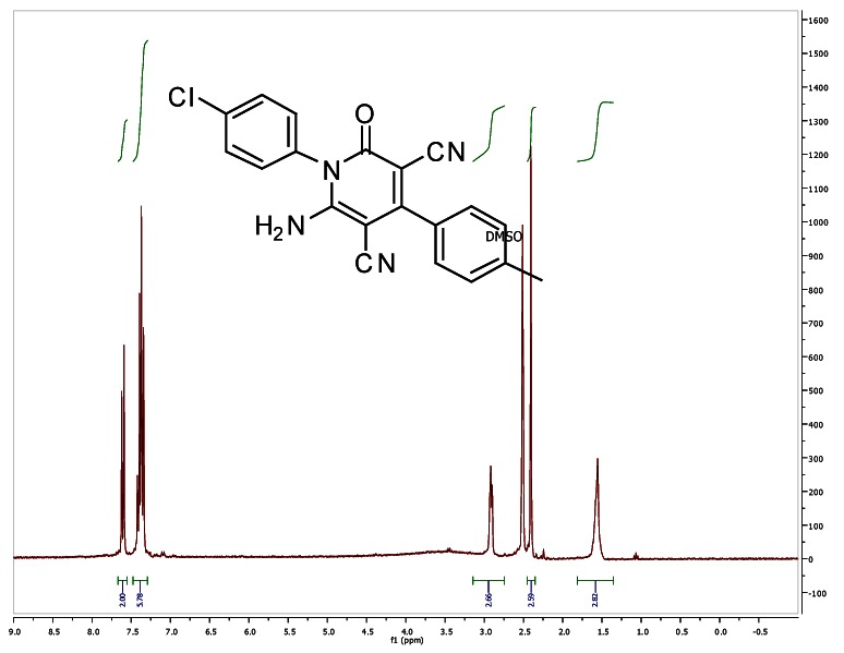
1H NMR (300 MHz, d6-DMSO) (Table 1, entry 2)

LC-MS

1H NMR (300 MHz, d6-DMSO) (Table 1, entry 3)

LC-MS
1H NMR (300 MHz, d6-DMSO) (Table 1, entry 4)

LC-MS
1H NMR (300 MHz, d6-DMSO) (Table 1, entry 5)

LC-MS

1H NMR (300 MHz, CD3CN) (Table 1, entry 6)

LC-MS
Appendix 5. Compounds Submitted to BioFocus
| BRD | SID | CID | P/A | MLSID | ML |
|---|---|---|---|---|---|
| BRD-K50776152-001-07-0 | 87556782 | 824820 | Probe | MLS002699917 | ML160 |
| BRD-K12621773-001-01-7 | 87556783 | 44629412 | Analog | MLS002699918 | N/A |
| BRD-K12168891-001-05-7 | 87556784 | 1034641 | Analog | MLS002699919 | N/A |
| BRD-K12238169-001-01-2 | 87556786 | 12425537 | Analog | MLS002699920 | N/A |
| BRD-K84855052-001-01-6 | 87556787 | 44629408 | Analog | MLS002699921 | N/A |
| BRD-K29715937-001-02-9 | 87556785 | 1034643 | Analog | MLS002699923 | N/A |
Acknowledgements
The authors thank Patti Aha, Tyler Aldredge, Frank An, PJ Aspesi, Doug Barker, Jason Burbank, Josh Bittker, Dave DeCaprio, Melanie de Silva, Karen Emmith, Robert Gould, Mary Pat Happ, Kate Hartland, Tom Hasaka, Stephen Johnston, Corrie Lade, Hanh Le, John McGrath, Chris Moore, Evan Mulligan, Myra O’Leary, Gil Walzer, Greg Wendel, and other members of BIPDeC for their assistance with this project. The authors also acknowledge and thank BloodSource (CA), Rhode Island Blood Center, Blood Center of New Jersey, and Blood Center of Wisconsin for supplying platelets for the assays performed at BIPDeC.
- PMCPubMed Central citations
- PubChem BioAssay for Chemical ProbePubChem BioAssay records reporting screening data for the development of the chemical probe(s) described in this book chapter
- PubChem SubstanceRelated PubChem Substances
- PubMedLinks to PubMed
- Review Chemical Genetic Analysis of Platelet Granule Secretion-Probe 3.[Probe Reports from the NIH Mol...]Review Chemical Genetic Analysis of Platelet Granule Secretion-Probe 3.VerPlank L, Dockendorff C, Negri J, Perez JR, Dilks J, MacPherson L, Palmer M, Flaumenhaft R, Schreiber SL. Probe Reports from the NIH Molecular Libraries Program. 2010
- Review Chemical Genetic Analysis of Platelet Granule Secretion-Probe 1.[Probe Reports from the NIH Mol...]Review Chemical Genetic Analysis of Platelet Granule Secretion-Probe 1.VerPlank L, Dockendorff C, Negri J, Perez JR, Dilks J, MacPherson L, Palmer M, Flaumenhaft R, Schreiber SL. Probe Reports from the NIH Molecular Libraries Program. 2010
- Acid sphingomyelinase regulates platelet cell membrane scrambling, secretion, and thrombus formation.[Arterioscler Thromb Vasc Biol....]Acid sphingomyelinase regulates platelet cell membrane scrambling, secretion, and thrombus formation.Münzer P, Borst O, Walker B, Schmid E, Feijge MA, Cosemans JM, Chatterjee M, Schmidt EM, Schmidt S, Towhid ST, et al. Arterioscler Thromb Vasc Biol. 2014 Jan; 34(1):61-71. Epub 2013 Nov 14.
- Protease-activated receptors differentially regulate human platelet activation through a phosphatidic acid-dependent pathway.[Mol Pharmacol. 2007]Protease-activated receptors differentially regulate human platelet activation through a phosphatidic acid-dependent pathway.Holinstat M, Voss B, Bilodeau ML, Hamm HE. Mol Pharmacol. 2007 Mar; 71(3):686-94. Epub 2006 Dec 6.
- Platelets possess and require an active protein palmitoylation pathway for agonist-mediated activation and in vivo thrombus formation.[Arterioscler Thromb Vasc Biol....]Platelets possess and require an active protein palmitoylation pathway for agonist-mediated activation and in vivo thrombus formation.Sim DS, Dilks JR, Flaumenhaft R. Arterioscler Thromb Vasc Biol. 2007 Jun; 27(6):1478-85. Epub 2007 Feb 15.
- Chemical Genetic Analysis of Platelet Granule Secretion-Probe 2 - Probe Reports ...Chemical Genetic Analysis of Platelet Granule Secretion-Probe 2 - Probe Reports from the NIH Molecular Libraries Program
Your browsing activity is empty.
Activity recording is turned off.
See more...