U.S. flag

An official website of the United States government

NCBI Bookshelf. A service of the National Library of Medicine, National Institutes of Health.

Adam MP, Feldman J, Mirzaa GM, et al., editors. GeneReviews® [Internet]. Seattle (WA): University of Washington, Seattle; 1993-2024.

Cover of GeneReviews®

GeneReviews® [Internet].

Show details

APC-Associated Polyposis Conditions

, MD, , MD, , MS, CGC, and , MD, MS.

Author Information and Affiliations

Initial Posting: ; Last Update: May 12, 2022.

Estimated reading time: 54 minutes

Summary

Clinical characteristics.

APC-associated polyposis conditions include (classic or attenuated) familial adenomatous polyposis (FAP) and gastric adenocarcinoma and proximal polyposis of the stomach (GAPPS).

  • FAP is a colorectal cancer (CRC) predisposition syndrome that can manifest in either classic or attenuated form. Classic FAP is characterized by hundreds to thousands of adenomatous colonic polyps, beginning on average at age 16 years (range 7-36 years).
    For those with the classic form of FAP, 95% of individuals have polyps by age 35 years; CRC is inevitable without colectomy. The mean age of CRC diagnosis in untreated individuals is 39 years (range 34-43 years). The attenuated form is characterized by multiple colonic polyps (average of 30), more proximally located polyps, and a diagnosis of CRC at a later age than in classic FAP.
    For those with an attenuated form, there is a 70% lifetime risk of CRC and the mean age of diagnosis is 50-55 years. Extracolonic manifestations are variably present and include polyps of the stomach and duodenum, osteomas, dental abnormalities, congenital hypertrophy of the retinal pigment epithelium (CHRPE), benign cutaneous lesions, desmoid tumors, adrenal masses, and other associated cancers.
  • GAPPS is characterized by proximal gastric polyposis, increased risk of gastric adenocarcinoma, and no duodenal or colonic involvement in most individuals reported.

Diagnosis/testing.

The diagnosis of an APC-associated polyposis condition is established by identification of a heterozygous germline pathogenic variant in APC.

Management.

Treatment of manifestations: Resection of all colonic polyps larger than 5 mm found on colonic surveillance. There is an absolute indication for colectomy when CRC is diagnosed or suspected, or when there are significant symptoms (e.g., bleeding, obstruction). Relative indications for colectomy include presence of multiple adenomas larger than 10 mm that cannot be reasonably removed endoscopically, a significant increase in adenoma number between surveillance exams, presence of adenomas with high-grade dysplasia, or inability to adequately survey the colon (e.g., due to innumerable diminutive adenomas or limited access to or compliance with colonoscopy). Endoscopic or surgical removal of duodenal adenomas is considered if polyps exhibit villous change or severe dysplasia, exceed 1 cm in diameter, or exhibit advanced stage using Spigelman scoring system. Gastrectomy is considered if advanced gastric neoplasia is found on upper endoscopy. Osteomas may be removed for cosmetic reasons. Desmoid tumors may be surgically excised or treated with nonsteroidal anti-inflammatory drugs (NSAIDs), anti-estrogens, cytotoxic chemotherapy, and/or radiation if at advanced stage. Standard treatment when needed for adrenal masses and thyroid carcinoma. Several studies have shown that NSAIDs and erlotinib have caused regression of adenomas and decreased the polyp burden in individuals with FAP, though there are currently no FDA-approved chemopreventive agents for FAP, given an unclear effect on subsequent cancer risk.

Prevention of primary manifestations: Colectomy to reduce the risk for CRC in individuals with classic FAP. For individuals with attenuated FAP, colectomy may be necessary, but in approximately one third of individuals, the colonic polyps are limited enough in number that surveillance with periodic colonoscopic polypectomy is sufficient to prevent CRC. It is currently unknown if prophylactic gastrectomy should be considered in individuals with GAPPS.

Surveillance: Colorectal screening by colonoscopy every one to two years beginning at age ten to 15 years for classic FAP and in late adolescence for attenuated FAP; esophagogastroduodenoscopy with visualization of the ampulla of Vater by age 20 to 25 years or prior to colon surgery, with consideration of complete small bowel visualization in the setting of advanced Spigelman stage. Annual thyroid palpation, thyroid ultrasound, neurologic examination, and abdominal examination (for desmoids). Liver palpation, liver ultrasound, and measurement of serum alpha-fetoprotein every three to six months until age five years for hepatoblastoma. The efficacy of screening for gastric cancer in individuals with GAPPS is currently unknown.

Agents/circumstances to avoid: Multistage surgeries in those at high risk for desmoids; total colectomy with ileal pouch anal anastomosis in women prior to childbearing.

Evaluation of relatives at risk: Molecular genetic testing for early identification of at-risk family members improves diagnostic certainty and reduces the need for costly screening procedures in those at-risk family members who have not inherited the pathogenic variant.

Genetic counseling.

APC-associated polyposis conditions are inherited in an autosomal dominant manner. Approximately 75%-80% of individuals with an APC-associated polyposis condition have an affected parent. Offspring of an affected individual are at a 50% risk of inheriting the pathogenic variant in APC. Prenatal testing and preimplantation genetic testing are possible if a pathogenic variant has been identified in an affected family member.

GeneReview Scope

APC-Associated Polyposis Conditions: Included Phenotypes 1
  • Familial adenomatous polyposis (FAP)
  • Attenuated FAP
  • Gastric adenocarcinoma and proximal polyposis of the stomach (GAPPS)

For synonyms and outdated names see Nomenclature.

1.

For other genetic causes of these phenotypes see Differential Diagnosis.

Diagnosis

Suggestive Findings

The National Comprehensive Cancer Network (NCCN) has published an algorithm for consideration of the diagnosis of both familial adenomatous polyposis (FAP) and attenuated FAP [Weiss et al 2021] (full text). These guidelines include recommendations for genetic testing of APC. Consensus guidelines specific for gastric adenocarcinoma and proximal polyposis of the stomach (GAPPS) are not yet available.

According to the NCCN guidelines, an APC-associated polyposis condition should be suspected in individuals with any of the following clinical features:

  • Multiple colorectal adenomatous polyps (at least 10-20 cumulative)
  • Family history of multiple colorectal adenomatous polyps (>10 in a single individual, or fewer if >1 relative has multiple polyps, especially if diagnosed at a young age), known APC pathogenic variant, and/or extracolonic features of APC-associated polyposis conditions
  • Hepatoblastoma
  • Multifocal/bilateral congenital hypertrophy of the retinal pigment epithelium (CHRPE)
  • Desmoid tumor
  • Cribriform-morular variant of papillary thyroid cancer

Additional features suggestive of an APC-associated polyposis condition include early-onset colorectal cancer with few to no adenomatous polyps, dental abnormalities (e.g., supernumerary teeth), osteomas, odontomas, epidermoid cysts, duodenal adenomas and cancer, gastric fundic gland polyposis, gastric cancer, pancreatic cancer, small bowel carcinoma, and/or medulloblastoma.

Establishing the Diagnosis

The diagnosis of an APC-associated polyposis condition is established by identification of a heterozygous germline pathogenic (or likely pathogenic) variant in APC (see Table 1).

Note: (1) Per ACMG/AMP variant interpretation guidelines, the terms "pathogenic variants" and "likely pathogenic variants" are synonymous in a clinical setting, meaning that both are considered diagnostic and both can be used for clinical decision making [Richards et al 2015]. Reference to "pathogenic variants" in this section is understood to include any likely pathogenic variants. (2) Identification of a heterozygous APC variant of uncertain significance does not establish or rule out the diagnosis.

The diagnosis of classic FAP is considered in an individual with a heterozygous germline pathogenic variant in APC identified by molecular genetic testing AND:

  • ≥100 colorectal adenomatous polyps (individuals at younger ages or those with colectomies may have <100 colorectal adenomatous polyps); OR
  • Multiple but <100 colorectal adenomatous polyps and a relative with confirmed classic FAP.

The diagnosis of attenuated FAP is considered in an individual with a heterozygous germline pathogenic variant in APC identified by molecular genetic testing AND:

  • A relative with confirmed attenuated FAP; AND/OR
  • <100 colorectal adenomatous polyps; OR
  • >100 colorectal adenomatous polyps at an advanced age (>age 40 years).

The diagnosis of GAPPS is considered in an individual with a heterozygous germline pathogenic variant in APC promotor 1B identified by molecular genetic testing AND:

  • Gastric polyps restricted to the body and fundus;
  • >100 polyps in the proximal stomach or >30 polyps in a first-degree relative of an individual with GAPPS;
  • Predominantly fundic gland polyps (FGPs) and some gastric adenomas; some having regions of dysplasia (or a family member with either dysplastic FGPs or gastric adenocarcinoma) [de Boer et al 2018];
  • No evidence of colorectal or duodenal polyposis.

Molecular Genetic Testing

  • Molecular genetic testing approaches can include single-gene testing and use of a multigene panel:
  • Single-gene testing. Testing should include both sequencing and deletion/duplication analysis of APC. Deletion/duplication testing should also include analysis of APC regulatory regions (specifically promoter 1B) if an APC pathogenic variant is not identified with initial testing.
    Note: APC sequence and deletion/duplication analysis on adenoma tissue can be considered in those with suspected APC-associated polyposis and negative germline testing. Identification of the same APC pathogenic variant in multiple adenomas in an individual with negative germline testing is consistent with somatic mosaicism [Aretz et al 2007]. Approximately 20% of simplex cases are somatic mosaic for an APC pathogenic variant.
  • A multigene panel that includes APC and other genes of interest (see Differential Diagnosis) may also be considered. Note: (1) The genes included in the panel and the diagnostic sensitivity of the testing used for each gene vary by laboratory and are likely to change over time. (2) Some multigene panels may include genes not associated with the condition discussed in this GeneReview; thus, clinicians need to determine which multigene panel is most likely to identify the genetic cause of the condition while limiting identification of variants of uncertain significance and pathogenic variants in genes that do not explain the underlying phenotype. (3) In some laboratories, panel options may include a custom laboratory-designed panel and/or custom phenotype-focused exome analysis that includes genes specified by the clinician. (4) Methods used in a panel may include sequence analysis, deletion/duplication analysis, and/or other non-sequencing-based tests.
    For an introduction to multigene panels click here. More detailed information for clinicians ordering genetic tests can be found here.

Table 1.

Molecular Genetic Testing Used in APC-Associated Polyposis Conditions

Gene 1MethodProportion of Probands with a Pathogenic Variant 2 Detectable by Method
APC Sequence analysis 3≤90% 4, 5
Gene-targeted deletion/duplication analysis 6~8%-12% 4
1.

See Table A. Genes and Databases for chromosome locus and protein.

2.

See Molecular Genetics for information on variants detected in this gene.

3.

Sequence analysis detects variants that are benign, likely benign, of uncertain significance, likely pathogenic, or pathogenic. Variants may include small intragenic deletions/insertions and missense, nonsense, and splice site variants; typically, exon or whole-gene deletions/duplications are not detected. For issues to consider in interpretation of sequence analysis results, click here.

4.

Data derived from the subscription-based professional view of Human Gene Mutation Database [Stenson et al 2020]

5.

Approximately 20% of simplex cases have somatic mosaicism [Hes et al 2008], and molecular genetic testing of DNA extracted from lymphocytes may fail to detect the APC pathogenic variant [Aretz et al 2007, Hes et al 2008].

6.

Gene-targeted deletion/duplication analysis detects intragenic deletions or duplications. Methods used may include a range of techniques such as quantitative PCR, long-range PCR, multiplex ligation-dependent probe amplification (MLPA), and a gene-targeted microarray designed to detect single-exon deletions or duplications. Large deletion/duplication testing should also include analysis of APC regulatory regions (specifically promoter 1B) [Rohlin et al 2011].

Clinical Characteristics

Clinical Description

APC-associated polyposis conditions include classic familial adenomatous polyposis (FAP), attenuated FAP, and gastric adenocarcinoma and proximal polyposis of the stomach (GAPPS).

FAP

In individuals with classic FAP, colorectal adenomatous polyps begin to appear in the second and third decade; the average age of polyp diagnosis is 16 years (range 7-36 years) [Petersen et al 1991]. By age 35 years, 95% of individuals with FAP have polyps. Once they appear, the polyps rapidly increase in number; when colonic expression is fully developed, hundreds to thousands of colonic adenomatous polyps are typically observed. Without colectomy, colorectal cancer (CRC) is essentially inevitable. The average age of CRC diagnosis in untreated individuals is 39 years (range 34-43 years); 7% of untreated individuals with FAP develop CRC by age 21 years, 87% by 45 years, and 93% by 50 years. Although rare, asymptomatic individuals in their 50s have been reported. Inter- and intrafamilial phenotypic variability are common [Half et al 2009].

Attenuated FAP is characterized by fewer colonic polyps (average of 30 polyps) than classic FAP but a significant risk for CRC. According to the international collaborative study by Knudsen et al [2010], attenuated FAP was clinically defined as individuals with ≤100 colorectal adenomatous polyps at age ≥25 years. Polyps tend to occur more proximally in the colon than in classic FAP. The average age of CRC diagnosis in individuals with attenuated FAP is 50 to 55 years – ten to 15 years later than in classic FAP, but earlier than in those with sporadically occurring CRC [Spirio et al 1993, Giardiello et al 1997]. The cumulative risk for CRC by age 80 years in attenuated FAP is estimated at 70% [Neklason et al 2008].

Other Features Variably Present in FAP

Table 2.

Lifetime Risk for Extracolonic Cancer in Familial Adenomatous Polyposis

SiteType of CancerLifetime Risk for Cancer
Small bowel: duodenum (most often periampullary region)Adenocarcinoma, carcinoma4%-12%
Small bowel: distal to the duodenumCarcinomaRare
PancreasAdenocarcinoma~1%
ThyroidPapillary thyroid carcinoma1%-12%
CNSUsually medulloblastoma~1%
LiverHepatoblastoma1.6%
Bile ductsAdenocarcinomaLow, but ↑
StomachAdenocarcinoma1.3% in Western cultures since 2016 [Mankaney et al 2017]

CNS = central nervous system

Small-bowel polyps and cancer. Adenomatous polyps of the duodenum, observed in 50%-90% of individuals with FAP, are commonly found in the second and third portions of the duodenum [Kadmon et al 2001] and to a variable degree in the jejunum or ileum [Koornstra 2012]. A classification system for duodenal polyps, based on number and size of polyps, histology, and degree of dysplasia, has been developed [Spigelman et al 1989]. No clear association between the number of colonic polyps and the number of upper gastrointestinal polyps has been identified [Kadmon et al 2001].

Adenomatous polyps of the periampullary region (including the duodenal papilla and ampulla of Vater), some of which can be endoscopically subtle or invisible, are seen in at least 50% of individuals with FAP, [Mehta et al 2021]. Polyps in this area can cause obstruction of the pancreatic duct resulting in pancreatitis or biliary obstruction, both of which occur at increased frequency in individuals with FAP. These polyps are often small and require either a distal attachment cap-assisted gastroscope or side-viewing endoscope for visualization [Yang et al 2020]. Some theorize that pancreaticobiliary secretions (e.g., bile) affect the development of adenomas [Wallace & Phillips 1998] and may account for the observed increased risk for malignancy of polyps in the periampullary region [Kadmon et al 2001].

The lifetime risk for small bowel malignancy is 4%-12%, with the large majority occurring in the duodenum. Duodenal adenocarcinoma occurs most commonly in the periampullary area. It has been reported to occur between ages 17 and 81 years, with the mean age of diagnosis between 45 and 52 years [Wallace & Phillips 1998, Kadmon et al 2001]. Small-bowel cancer distal to the duodenum occurs but is rare. Ruys et al [2010] identified only 17 reported instances of jejunal carcinoma and three reports of ileal carcinoma in individuals with FAP.

Pancreatic cancer. While limited data exist, one study of 197 families with FAP revealed a relative risk for pancreatic cancer of 4.5 in individuals with FAP and their at-risk relatives compared to the general population. Giardiello et al [1993b] estimated the pancreatic cancer risk to age 80 years in individuals with FAP at 1%. Since that report, there have only been isolated case reports of pancreatic cancer in individuals with FAP [Moussata et al 2015].

Thyroid cancer and benign thyroid disease. A high degree of variability in the frequency of thyroid cancer is reported in individuals with FAP. Various retrospective reviews have reported a prevalence of 0.4% to 2.6%, whereas prospective studies have found a higher prevalence of 2.6% to 11.8% [Cetta 2015, Chenbhanich et al 2019]. There is a striking female-to-male ratio of 80 to 1 in FAP, and more than 80% of individuals are diagnosed between ages 18 and 35 years [Cetta 2015]. Papillary histology predominates and may have a cribriform pattern [Chenbhanich et al 2019]. A rare subtype of papillary thyroid carcinoma, cribriform-morular variant, is typically associated with FAP [Pradhan et al 2015], although it can also occur as a sporadic cancer.

Data on the rate of benign thyroid disease in individuals with FAP are limited. In a systematic review and meta-analysis, 6.9% of individuals with FAP had benign endocrinologic thyroid disease (hypothyroidism, goiter, and/or thyroiditis) and 48.8% had benign thyroid nodules [Chenbhanich et al 2019]. Significant heterogeneity among studies likely contributes to the discrepancies in the reported rates of thyroid disease. Familial occurrence and a female preponderance have been observed.

Central nervous system (CNS). Medulloblastoma is the most common CNS tumor identified in individuals with FAP. The risk for CNS tumors is substantially increased in persons with FAP, although the absolute risk is only approximately 1% [Attard et al 2007].

Hepatoblastoma. The risk for hepatoblastoma in individuals with FAP is 750 to 7,500 times higher than in the general population, although the absolute risk is estimated at less than 2%. The majority of hepatoblastomas occur before age three years [Aretz et al 2007].

Gastric polyps and cancer. The risk for both fundic gland and adenomatous polyps of the stomach is increased in FAP. Gastric fundic gland polyps (FGPs) are benign neoplasms located in the fundus and body of the stomach; some authors classify them as hamartomatous, but this classification is under debate. FGPs occur in approximately half of individuals with FAP and undergo dysplastic change more commonly than sporadic FGPs [Bianchi et al 2008]. Adenomatous polyps (e.g., pyloric gland adenomas, tubular adenomas) and hyperplastic polyps can be seen in individuals with FAP.

The risk for gastric cancer in individuals with FAP living in Western cultures is low, but has risen in recent years [Mankaney et al 2017]. The rates of gastric cancer in persons of Japanese and Korean ancestry with FAP may be tenfold higher than the general population [Garrean et al 2008]. Gastric adenocarcinoma is believed to arise most often from adenomas but may also develop from FGPs [Attard et al 2001]. Classification systems have been proposed to assist in endoscopic identification of high-risk polyps [Mankaney et al 2020].

Non-Malignant Extraintestinal Manifestations of FAP

Osteomas occur in about 60%-80% of individuals with FAP. They are bony growths found most commonly on the skull and mandible, but they may occur in any bone of the body. Osteomas do not usually cause clinical problems and do not become malignant; they may appear in children prior to the development of colonic polyps [Septer et al 2018].

Dental abnormalities. Unerupted teeth, congenital absence of one or more teeth, supernumerary teeth, dentigerous cysts (an odontogenic cyst associated with the crown of an unerupted tooth), and odontomas have been reported in approximately 30%-75% of individuals with FAP compared to 1%-2% of the general population [Septer et al 2018].

Congenital hypertrophy of the retinal pigment epithelium (CHRPE) refers to discrete, flat, pigmented lesions of the retina that are not age dependent and do not cause clinical problems. CHRPE is reported to occur in up to 80% of individuals with FAP. Visualization of CHRPE may require examination of the ocular fundus with an indirect ophthalmoscope through a dilated pupil. Observation of multiple or bilateral CHRPE may be an indication that an at-risk family member has inherited FAP, whereas isolated lesions can be seen in the general population [Rehan & Aye 2020].

Benign cutaneous lesions include epidermoid cysts and fibromas that may be found on any part of the body, including the face. They are mainly of cosmetic concern, as they do not appear to have malignant potential. Multiple pilomatricomas (benign tumors of the hair follicles), although rare, have also been reported [Ciriacks et al 2020].

Desmoid tumors develop in approximately 10%-30% of individuals with FAP [Nieuwenhuis et al 2011b, Sinha et al 2011]. The risk for desmoid tumors in individuals with FAP is more than 800 times the risk in the general population. At least 7.5% of desmoid type fibromatoses are found in people with FAP [Nieuwenhuis et al 2011a]. These poorly understood, benign fibrous tumors are clonal proliferations of myofibroblasts that are locally invasive but do not metastasize. A pathologically distinct fibromatous lesion called a Gardner-associated fibroma is hypothesized to be a precursor lesion [Wehrli et al 2001].

The incidence of desmoid tumors in individuals with FAP is highest in the second and third decades of life, with 80% occurring by age 40 years [Sinha et al 2011]. Approximately 65% of desmoid tumors in individuals with FAP occur within the abdomen or in the abdominal wall [Sinha et al 2011], although they can occur along the axial skeleton or extremities as well [Escobar et al 2012]. Desmoid tumors may compress abdominal organs or complicate abdominal surgery. About 5% of individuals with FAP experience morbidity and/or mortality from desmoid tumors, with the highest mortality rate reported for intra-abdominal tumors [Sinha et al 2011]. Abdominal desmoid tumors may occur spontaneously or following abdominal surgery [Bertario et al 2001]. The effect of pregnancy on desmoid tumor growth or development is unknown [Sinha et al 2011]. Independent predictors for desmoid tumor development include: an APC pathogenic variant 3' of codon 1399, family history of desmoid tumors, female gender, and previous abdominal surgery [Sinha et al 2011]. An APC pathogenic variant 5' of codon region 543-713 has also been associated with desmoid tumors [Slowik et al 2015]. Positive family history of desmoid tumor was associated with the highest magnitude of risk; having a first-degree relative with a desmoid tumor was associated with a seven-fold increase in risk [Sinha et al 2011].

Desmoid tumors are best evaluated by MRI or CT scan [Escobar et al 2012]. A CT scoring system for desmoid tumors in FAP has been developed [Middleton et al 2003].

Adrenal masses are two to four times more prevalent in individuals with FAP than in the general population [Rekik et al 2010]. Adrenal masses are found in 1%-3% of the general population compared to 7.4%-16% of individuals with FAP [Marchesa et al 1997, Shiroky et al 2018]. A prospective study of 107 individuals with FAP found 13% with an adrenal mass ≥1.0 cm on abdominal CT scan [Smith et al 2000b]. Most of the masses are asymptomatic adenomas found incidentally, although functional lesions and carcinomas do occur [Marchesa et al 1997, Rekik et al 2010].

GAPPS

GAPPS is characterized by proximal gastric FGPs and intestinal-type gastric adenocarcinoma, typically without significant duodenal or colorectal polyposis [Worthley et al 2012]. Individuals with GAPPS have a 13%-25% lifetime risk for gastric carcinoma [Kim et al 2022].

Genotype-Phenotype Correlations

Although inter- and intrafamilial variability is common in FAP, there are some reported genotype-phenotype correlations (see Table 3). However, surveillance and prophylactic surgery should be tailored to the affected individual's phenotype and not based solely on genotype. Some studies report wide phenotypic variability in individuals with the same variant.

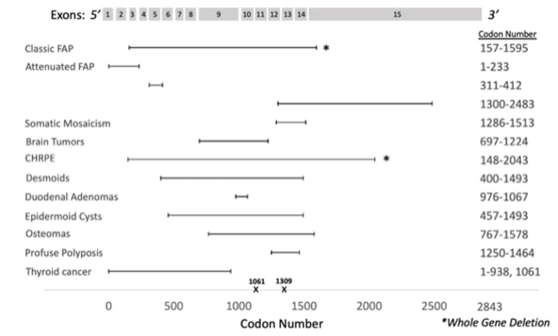
Of individuals with phenotypic criteria of attenuated FAP, 78% had a pathogenic variant in one of three regions: the 5' end of APC (codon 1-233), the alternative spliced region of exon 9 (codon 311-412), or 3' of codon 1595. However, genotype does not always predict colorectal phenotype; of individuals with a pathogenic variant in one of these three regions typically associated with attenuated FAP, 65% had ≤100 colorectal adenomas and 35% had classic FAP and had undergone colectomy. The median age of colectomy was 18 years [Anele et al 2022].

Figure 1.

Figure 1.

Schematic representation of APC

APC consists of 15 exons and 2843 codons. Most germline variants are located in the 5' half of APC, particularly in codons 1061 and 1309. Germline variants in the central part of the gene are often associated with classic FAP. Germline variants between codons 1250 and 1464 are associated with profuse polyposis, whereas variants in the 5' or 3' ends cause attenuated FAP.

Table 3.

Genotype-Phenotype Correlations in APC-Associated Conditions

PhenotypeLocation/Type of APC Pathogenic VariantCommentsReference(s)
FAP (classic form) Whole-gene deletion Quadri et al [2015]
Attenuated FAP
  • 5' end (codons 1-233) 1
  • Distal 3' end (1300-2843; esp 3' of codon 1585)
  • Exon 9 (codons 311-412)
Extraintestinal manifestations (e.g., CHRPE, desmoid tumors) are rare.Sieber et al [2006], Anele et al [2022]
GAPPS APC promoter 1B
Somatic mosaicism Codons 1286-1513 (most common)Colon phenotype:
  • 65% attenuated FAP
  • 30% FAP
  • 5% no adenomas
Friedl & Aretz [2005], Hes et al [2008], Jansen & Goel [2020]
Brain tumors Codons 697-1224
  • 3x ↑ risk brain tumor
  • 13x ↑ risk medulloblastoma
Attard et al [2007]
CHRPE Codons 148-2043 Burger et al [2011]
Codons 311-1444See footnote 2.
Whole-gene deletion Aretz et al [2005]
Desmoids Codons 1395-1493Odds ratio: 4.37 Sinha et al [2011]
5' of codon 400Incidence: 14.9% Church et al [2015]
Codons 401-1400Incidence: 23.3%
3' of codon 1400Incidence: 37.1%
Duodenal adenomas Codons 976-10674x ↑ risk Bertario et al [2003]
Epidermoid cysts Codons 457-1493 Dinarvand et al [2019]
Osteomas Codons 767-1578
Profuse polyposis Codons 1250-1464Average of 5,000 polyps D'Elia et al [2018]
Thyroid cancer 5' end (codon 1061 or proximal to codon 938) Chenbhanich et al [2019]

CHRPE = congenital hypertrophy of the retinal pigment epithelium; FAP = familial adenomatous polyposis; GAPPS = gastric adenocarcinoma and proximal polyposis of the stomach

1.

Variants 5' of codon 233 are the most commonly described variants associated with attenuated FAP [Knudsen et al 2003].

2.

Data derived from the subscription-based professional view of Human Gene Mutation Database [Stenson et al 2020]

Penetrance

In FAP, the penetrance is 100%.

In attenuated FAP, the penetrance of colonic polyposis is less well understood, although the estimate of CRC risk by age 80 years is approximately 70% [Neklason et al 2008].

In GAPPS, the penetrance of FGPs and/or intestinal-type gastric adenocarcinoma is unknown.

Nomenclature

FAP is often referred to as classic FAP when more than 100 colorectal polyps are present. Classic FAP and FAP may be used interchangeably.

Adenomatous polyposis coli (APC) was used historically to refer to FAP; APC now refers to the relevant gene.

A variety of terms have been used to describe individuals with an APC-associated polyposis condition: FAP, attenuated FAP, Gardner syndrome, Turcot syndrome, and gastric adenocarcinoma and proximal polyposis of the stomach (GAPPS). The clinical features associated with these phenotypes are addressed in this GeneReview; however, all are now genetically defined as caused by pathogenic variants in APC. Terms such as Gardner syndrome and Turcot syndrome are of historical interest and should not be used, as both are now known to be part of the FAP spectrum.

Prevalence

Estimates of the prevalence of FAP vary from 1:6,850 to 1:31,250 live births (2.29-3.2 cases per 100,000 individuals) [Half et al 2009, Scheuner et al 2010]. The frequency is fairly constant throughout the world, with men and women being equally affected. Attenuated FAP is likely underdiagnosed, given the lower number of colonic polyps and lower risk for CRC compared to FAP [Neklason et al 2008].

The prevalence of GAPPS is currently unknown.

APC-associated polyposis conditions historically accounted for about 0.5% of all CRC diagnoses; this figure is declining as more at-risk family members undergo successful treatment following early polyp detection and prophylactic colectomy.

Differential Diagnosis

Hereditary Disorders to Consider in the Differential Diagnosis

APC-associated polyposis conditions may be distinguished from other inherited colorectal cancer conditions and other gastrointestinal polyposis syndromes (see Table 4) by molecular genetic testing, histopathologic findings, and phenotypic characteristics.

Table 4.

Selected Hereditary Polyposis and Colorectal Cancer Syndromes in the Differential Diagnosis of APC-Associated Polyposis Conditions

Gene(s) / Genetic MechanismDisorderMOIComment
15q15.3q22.1 duplication 1
BMPR1A
SMAD4
Hereditary mixed polyposis syndrome (HMPS) (OMIM 601228)ADAssoc w/↑ risk for CRC & multiple different types of colorectal polyps. Characteristic lesions are mixed juvenile-adenomatous colon polyps. Adenomas, hyperplastic serrated adenomas, & mixed hyperplastic-adenomatous polyps may also occur.
AXIN2 AXIN2-assoc polyposis (oligodontia-CRC syndrome) (OMIM 608615)ADEctodermal dysplasia
BMPR1A
SMAD4
Juvenile polyposis syndrome (JPS)ADCharacterized by predisposition for hamartomatous polyps, which is often the distinguishing feature between FAP & JPS. Hamartomatous polyps occur in the GI tract – specifically stomach, small intestine, colon, & rectum. Most persons w/JPS have some polyps by age 20 yrs. Most juvenile polyps are benign, but malignant transformation can occur.
EPCAM
MLH1
MSH2
MSH6
PMS2
Lynch syndrome (hereditary non-polyposis colon cancer)ADIt may be difficult to distinguish Lynch syndrome from attenuated FAP in persons w/early-onset CRC & few adenomatous colonic polyps. Family history of extracolonic cancers & manifestations, MSI testing, &/or IHC testing on tumor tissue may be helpful in distinguishing the 2 disorders.
MLH1
MSH2
MSH6
PMS2
Constitutional mismatch repair deficiency (CMMRD) (See Lynch syndrome.)ARAffected persons frequently have brain tumors, hematologic malignancies, CRC, &/or other Lynch syndrome cancers in childhood. Café au lait macules &/or axillary/inguinal freckling are seen in most persons; multiple colorectal adenomas mimicking attenuated FAP may also be present.
MSH3 MSH3-assoc polyposis (OMIM 617100)ARColorectal & duodenal adenomas, CRC, gastric cancer, & early-onset astrocytoma
MUTYH MUTYH polyposis (MAP)ARAssoc w/predisposition to multiple adenomas or polyposis coli. The colonic phenotype of MAP can be similar to attenuated FAP. If an APC pathogenic variant is not identified in a person w/colonic polyposis, molecular genetic testing of MUTYH should be considered.
NF1 Neurofibromatosis type 1 (NF1)ADPersons w/NF1 may exhibit multiple intestinal polypoid neurofibromas or ganglioneuromas in small bowel, stomach, & colon.
NTHL1 NTHL1 tumor syndrome (NTHL1-assoc polyposis)ARCharacterized by ↑ lifetime risk for CRC, breast cancer, & colorectal polyposis. Addl cancers incl endometrial, cervical, urothelial carcinoma of the bladder, meningioma, unspecified brain tumors, basal cell carcinoma, head & neck squamous cell carcinoma, & hematologic malignancies.
POLD1 POLD1-assoc polyposis (polymerase proofreading-assoc polyposis) (OMIM 612591)ADColorectal & duodenal adenomas; MDPL syndrome (mandibular hypoplasia, deafness, progeroid features & lipodystrophy)
POLE POLE-assoc polyposis (polymerase proofreading-assoc polyposis) (OMIM 615083)ADColorectal & duodenal adenomas
PTEN Cowden syndrome (CS) (See PTEN Hamartoma Tumor Syndrome.)ADCS is assoc w/multiple colorectal polyps, but (unlike APC-assoc polyposis conditions) hamartomatous polyps, juvenile polyps, lipomas, & ganglioneuromas predominate. CS is assoc w/↑ risk of CRC, but breast, thyroid, & endometrial cancer are more common.
STK11 Peutz-Jeghers syndrome (PJS)ADCharacterized by assoc of GI PJS-type polyps & mucocutaneous pigmentation, neither of which are seen in APC-assoc polyposis conditions. PJS polyps are often symptomatic & most prevalent in small intestine (jejunum, ileum, & duodenum in order of prevalence) but can occur elsewhere in GI tract.

AD = autosomal dominant; AR = autosomal recessive; CRC = colorectal cancer; FAP = familial adenomatous polyposis; GI = gastrointestinal; IHC = immunohistochemistry; MOI = mode of inheritance; MSI = microsatellite instability

1.

HMPS can be caused by either a BMPR1A pathogenic variant or a duplication of 15q15.3q22.1 that leads to increased expression of GREM1 [Jaeger et al 2012]. Some families with mixed hereditary polyposis syndrome have SMAD4 pathogenic variants [Valle et al 2019].

Acquired Conditions to be Considered in the Differential Diagnosis

Cronkhite-Canada syndrome is characterized by generalized gastrointestinal hamartomatous polyposis, cutaneous hyperpigmentation, hair loss, and nail atrophy.

Nodular lymphoid hyperplasia, a lymphoproliferative disorder resulting in hyperplastic lymphoid nodules in the small bowel, stomach, and colon, may be associated with common variable immunodeficiency syndrome.

Lymphomatous polyposis is characterized by the occurrence of primary extranodal lymphomas in the gastrointestinal tract. Two types include multiple lymphomatous polyposis and Mediterranean-type lymphoma.

Inflammatory polyposis is characterized by acquired, non-neoplastic polyps associated with inflammatory bowel disease.

Sporadic colorectal tumors. The majority of colorectal tumors not known to be familial are associated with a somatic pathogenic variant in APC (see Genetically Related Disorders) [Lüchtenborg et al 2004, Christie et al 2013] that is believed to occur early in colorectal tumorigenesis [Christie et al 2013, Aghabozorgi et al 2019].

Therapy-associated polyposis has been reported as a possible cause of gastrointestinal polyposis [Biller et al 2020]. Thirty-four individuals treated at eight centers had gastrointestinal polyposis and a history of childhood or young-adult cancer treated with radiotherapy and/or chemotherapy at least ten years prior to identification of polyposis [Biller et al 2020]. However, it is unclear whether the chemotherapy and/or radiation caused the gastrointestinal polyposis or was coincidentally present in these individuals.

Other

Colonic adenomatous polyposis of unknown etiology. Many individuals with colonic adenomatous polyposis (≥10 cumulative colorectal polyps) will have uninformative germline molecular testing [Stanich et al 2019]. Note: The possibility of undetected APC somatic mosaicism should be considered in these individuals [Jansen & Goel 2020].

Serrated polyposis syndrome (previously termed hyperplastic polyposis) comprises multiple colorectal serrated polyps (hyperplastic polyps, sessile serrated adenomas/polyps, and traditional serrated adenomas). It is unknown whether this condition is inherited or acquired [Snover et al 2010]; pathogenic variants in RNF43 have been possibly implicated in a small subset of individuals [Yan et al 2017]. Although serrated polyps typically predominate, individuals with serrated polyposis frequently have multiple colorectal adenomas as well [Kalady et al 2011]. Individuals with serrated polyposis syndrome may also have a family history of colorectal cancer, although it is uncommon for more than one member of a family to meet the diagnostic criteria for serrated polyposis syndrome.

Management

Evaluations Following Initial Diagnosis

Individuals who are diagnosed with classic familial adenomatous polyposis (FAP) or attenuated FAP should be counseled about age-appropriate recommendations for surveillance and prevention of primary manifestations, in addition to treatment of manifestations, as outlined in this section. Currently, consensus management guidelines for gastric adenocarcinoma and proximal polyposis of the stomach (GAPPS) are unavailable.

Treatment of Manifestations

Practice parameters, including information on surgery, have been outlined by the following professional groups:

Colonic polyps. For individuals with FAP, endoscopic surveillance with colonoscopy is generally recommended to start between ages ten and 15 years with an interval of every one to two years [Weiss et al 2016, van Leerdam et al 2019, Yang et al 2020]. All polyps >5 mm should be resected. If the polyp burden is manageable and no definitive indications for surgery are noted, it is reasonable to delay colectomy and monitor with endoscopic surveillance [Ishikawa et al 2016]. Absolute indications for colectomy include documented or suspected colorectal cancer (CRC) or significant symptoms (e.g., obstruction, bleeding), although these are uncommon in the absence of cancer. Relative indications for colectomy include presence of multiple large adenomas (>10 mm) that cannot be reasonably managed by endoscopy, a significant increase in adenoma number between surveillance examinations, presence of adenomas with high-grade dysplasia, or inability to adequately survey the colon (e.g., due to innumerable diminutive adenomas or limited access or compliance with colonoscopy). In individuals age ten to 20 years in whom adenomas are <6 mm and without villous component or high-grade dysplasia, delay in colectomy may be considered to allow for physical and emotional maturity.

For individuals with an attenuated FAP phenotype, delaying the initiation of colonoscopy until late adolescence can be considered [Weiss et al 2016, van Leerdam et al 2019, Yang et al 2020]. Colonoscopic surveillance and polypectomy every one to two years can often be effective and delay or even prevent the need for colectomy [Knudsen et al 2010]. Approximately one third of individuals have colonic polyps that are limited enough in number that surveillance with periodic colonoscopic polypectomy is sufficient (see Surveillance) [Patel et al 2016].

Surgical options for colectomy include the following:

  • Total proctocolectomy with ileal pouch anal anastomosis (IPAA). This can be performed laparoscopically, laparoscopically-assisted, robotically, or open. The IPAA can be stapled, leaving 1-2 cm of anal transition epithelium and low rectal mucosa; or it can be hand-sewn after a complete anal mucosectomy. This is a multistage surgery.
  • Total colectomy with ileorectal anastomosis (IRA). This can also be performed with minimally invasive surgical techniques and is a single-stage surgery.
  • Total proctocolectomy with permanent ileostomy. This can also be performed with minimally invasive surgical techniques and is a single-stage surgery.

The choice of procedure depends on the clinical circumstances.

  • An IPAA is generally performed in individuals with FAP with a high rectal polyp burden (generally considered as >20 adenomas in the rectum or presence of advanced rectal neoplasia) or as a second procedure after IRA when rectal disease burden cannot be managed endoscopically [Warrier & Kalady 2012]. The advantage of this procedure is near-elimination of the risk for rectal cancer and relatively good preservation of bowel function. However, there may be an increased risk of bladder/sexual dysfunction compared to colectomy with IRA and functional results can be variable.
    A study of individuals with FAP and ileal pouches found that 57% had adenomatous polyps in the ileal pouch [Groves et al 2005].
  • An IRA is generally considered when the rectal polyp burden is minimal and deemed to be endoscopically manageable (usually in the setting of attenuated FAP). It is a technically straightforward procedure with low complication rates. It is usually associated with good functional outcome and minimizes risk of sexual or urinary dysfunction. When performed in appropriate individuals, the risk for rectal cancer or need for proctectomy after IRA is low [Church et al 2001]. This is not an optimal surgical choice if there is severe rectal disease or the individual cannot reliably undergo endoscopic surveillance of the remaining rectum postoperatively.
  • A total proctocolectomy with end ileostomy is rarely required unless a contraindication to IPAA is present (e.g., a mesenteric desmoid preventing a pouch from reaching pelvic floor, low rectal cancer invading pelvic floor, or individual preference due to poor sphincter control) and a proctocolectomy is necessary (due to rectal polyp/cancer burden).

Duodenal adenomas. Current guidelines recommend duodenal screening at age 20-25 years or earlier if colectomy is planned [van Leerdam et al 2019, Yang et al 2020, Weiss et al 2021]. This recommendation applies to all colon phenotypes and to both classic and attenuated FAP. Surveillance intervals are determined based on the Spigelman scoring system [Spigelman et al 1989], which incorporates duodenal polyp number, size, histology, and dysplasia grade to categorize affected individuals into five stages (see Table 5). Guidelines generally agree that for low-risk individuals (Stage 0 and I) a five-year interval for duodenal screening is appropriate, and for moderate-risk individuals (Stage II) a three-year interval is recommended. For higher-risk individuals (Stage III and IV) at least annual endoscopy is needed; for individuals with Stage IV there should be a consideration of surgical referral.

Table 5.

Spigelman Scoring System for Duodenal Adenomas in Familial Adenomatous Polyposis

Spigelman Scoring System
Criteria1 point2 points3 points
Polyp number1-45-20>20
Polyp size (mm)1-45-10>10
HistologyTubularTubulovillousVillous
DysplasiaMildModerateSevere

Stage 0 = 0 points

Stage I = 1-4 points

Stage II = 5-6 points

Stage III = 7-8 points

Stage IV = 9-12 points

Endoscopic or surgical removal of duodenal and/or ampullary adenomas is recommended by standard polypectomy techniques such as snaring and endoscopic mucosal resection. If there are too many polyps to remove, the focus should be on removing polyps >1 cm in size or those with concerning features. European Society of Gastrointestinal Endoscopy guidelines [van Leerdam et al 2019] caution against removing small polyps due to concern for fibrosis limiting future resection, but this has not been reported as a limiting factor in clinical practice.

Indications for surgery for advanced duodenal polyposis are Stage III with high-grade dysplasia, Stage IV disease, and malignancy. Surgical options for advanced duodenal polyposis include pancreaticoduodenectomy (Whipple procedure) and pancreas-sparing duodenectomy, which is a good option when the papilla is not involved and there is no suspicion for cancer. These surgeries have high associated morbidities and should be performed at high-volume centers and preferably by those with expertise in FAP.

Ampullary adenomas. Specific endoscopic techniques are needed to ensure adequate examination. This can be accomplished with a side-viewing duodenoscope or a clear-cap distal attachment to a forward-viewing gastroscope – approaches that have been found to be equivalent [Abdelhafez et al 2019]. Ampullary biopsy has been shown to be safe with a low risk for pancreatitis and there should be a low threshold to biopsy if there is a suspicion for an adenoma [Mehta et al 2021]. Guidelines vary on whether to include ampullary adenomas with duodenal polyposis in determining a surveillance interval; if a small ampullary adenoma is identified, most experts recommend repeat surveillance within three years. Although small ampullary adenomas can be monitored without resection, any adenomas >1 cm in size or with advanced histology should be resected. Ampullectomy carries a high rate of complications and should be performed by experienced providers [Roos et al 2021]. Recurrence after ampullectomy is common and close endoscopic surveillance is needed. Surgical considerations for ampullary adenomas are similar to those for advanced duodenal polyposis.

Gastric polyps. Gastric surveillance should be performed at the time of duodenal surveillance. Recommended guidelines for gastric surveillance are not as developed as those for duodenal polyposis, but with increasing incidence of gastric cancer in individuals with FAP these guidelines will likely evolve. Removal of all polyps that are concerning for adenomas / pyloric gland adenomas or advanced changes (dysplasia) is recommended along with random sampling of fundic gland polyps (FGPs). Some experts recommend using polyp number, size, histology, dysplasia, and other features to guide surveillance [Mankaney et al 2017]; endoscopic criteria (Surveillance for Pathology Associated with Cancer on Endoscopy; see Mankaney et al [2020]) can aid in optical diagnosis of high-risk pathology. If advanced neoplasia is noted on sampling, surgical gastrectomy should be considered.

Thyroid nodules and cancer. Treatment of thyroid nodules and papillary thyroid carcinoma, including the cribriform variant, is similar to that of sporadic disease [Abdullah Suhaimi et al 2015].

Osteomas may be removed for cosmetic reasons.

Desmoid tumors. Available treatments include surgical excision (associated with high rates of recurrence), nonsteroidal anti-inflammatory drugs (NSAIDs), anti-estrogens, cytotoxic chemotherapy, and radiation [Smith et al 2000a, Tonelli et al 2003, Gega et al 2006]. A review of desmoid treatments can be found in Guillem et al [2006] and the Desmoid Tumor Working Group [2020].

Adrenal tumors. Standard treatment is indicated as needed for adrenal masses.

Chemoprevention. There are currently no FDA-approved chemopreventive agents for FAP. Individuals interested in chemoprevention should be encouraged to enroll in an ongoing clinical trial (see Therapies Under Investigation). Note: The FDA has stated that changes in adenoma number and size are insufficient for approval and that clear evidence of clinical benefit is required. Cited examples of clinical benefit include decreased risk for CRC or reduced need for surgery; current trials are designed to address these endpoints.

NSAIDs. Non-placebo-controlled trials and observational studies on sulindac were initially promising, showing remarkable reduction in polyp size and number. However, these preliminary studies were limited in their design (non-placebo controlled; limited number of affected individuals; some individuals with only surveyable rectum). Several controlled trials subsequently confirmed a decrease in polyp burden during sulindac therapy [Labayle et al 1991, Giardiello et al 1993a, Nugent et al 1993]. However, rapid reappearance or increase in polyp number was observed after sulindac was discontinued [Labayle et al 1991, Giardiello et al 1993a]. A subsequent study designed to evaluate primary prevention of polyps in individuals with APC pathogenic variants showed a statistically nonsignificant trend toward benefit compared to placebo [Giardiello et al 2002].

The FDA initially approved celecoxib for FAP based on evidence of decreased colon polyp burden and size (as well as modest decrease in the duodenum) [Steinbach et al 2000, Phillips et al 2002]. However, due to cardiovascular and cerebrovascular safety concerns, FDA approval for celecoxib for FAP was withdrawn and rofecoxib was also taken off the market.

Aspirin has traditionally been shown to be of little or no benefit in FAP [Burn et al 2001, Ishikawa et al 2013], but a recent randomized trial showed a potential benefit in suppressing large polyps [Ishikawa et al 2021].

Interest in combination of NSAIDs with other drugs was raised when reports of sulindac plus difluoromethylornithine (DFMO) showed marked reduction in sporadic metachronous adenomas [Meyskens et al 2008]. In a randomized placebo-controlled study of 92 participants with FAP, sulindac plus erlotinib (an EGF receptor inhibitor) resulted in decreased duodenal polyp burden compared to placebo after six months of use [Samadder et al 2016]. Adverse events were common in the treatment group (87% experienced an acne-like rash), although serious adverse events were rare (2 participants) [Samadder et al 2016]. A secondary analysis of this trial also showed a decrease in colorectal polyp burden [Samadder et al 2018]. A criticism of this trial is that the endpoints are not clearly clinically meaningful. When celecoxib was compared to celecoxib plus DFMO, there was no significant difference in polyp burden within a defined endoscopic field (however, when more comprehensive video assessment was used, there was a decrease in polyp burden in the combination therapy group) [Lynch et al 2016]. More recently, a large trial did not show that disease progression was different with the combination of sulindac and eflornithine than with either drug separately [Burke et al 2020].

Note: NSAID use before colectomy remains experimental (see Therapies Under Investigation).

GAPPS. There are no current guidelines for management of GAPPS, but some experts have recommended gastroscopy starting at age 15 years with removal of all polyps that are concerning for adenomas / pyloric gland adenomas or advanced changes (dysplasia) along with random sampling of FGPs. If advanced neoplasia is noted on sampling, standard surgical gastrectomy should be considered [Author, personal communication].

Prevention of Primary Manifestations

FAP. Colectomy is advised to reduce the risk for CRC in classic FAP. For individuals with attenuated FAP, colectomy may be necessary, but in approximately one third of individuals, the colonic polyps are limited enough in number that surveillance with periodic colonoscopic polypectomy is sufficient to prevent CRC.

GAPPS. It is currently unknown if prophylactic gastrectomy should be considered in individuals with GAPPS. Some experts have recommended considering prophylactic gastrectomy at approximately age 30 years [Tacheci et al 2021].

Surveillance

Multiple professional societies have published guidelines based on the available evidence to date as well as expert consensus [Syngal et al 2015, Herzig et al 2017, Weiss et al 2021]. The following surveillance recommendations are based on these expert society guidelines.

Table 6.

Recommended Surveillance for Individuals with FAP

System/ConcernEvaluationFrequency/Comment
Colorectal
adenomatous
polyps
ColonoscopyEvery 1-2 yrs beginning: at age 10-15 yrs for classic FAP; in late adolescence for attenuated FAP
In persons who have undergone total colectomy w/IPAA: endoscopic surveillance of ileal pouchEvery 1-2 yrs
In persons who have undergone subtotal colectomy w/ileorectal anastomosis: surveillance of remaining rectum
  • Every 1-2 years or more frequently if high polyp burden
  • Note: Cancer may occur in remaining rectum, but risk is low. 1
In persons who have undergone total colectomy w/end ileostomy: ileoscopyEvery 1-2 yrs
Small-bowel
polyps & cancer
EGD w/complete visualization of ampulla of Vater (using duodenoscope or clear cap)Every 6 mos-5 yrs depending on duodenal adenoma burden 2 beginning at age 20-25 yrs or prior to colectomy
Consider complete small bowel visualization by video capsule endoscopy or CT/MR enterography esp if duodenal polyposis is advanced (based on Spigelman scoring system).
Thyroid cancer
  • Palpation of thyroid
  • Thyroid ultrasound
  • Fine-needle aspiration if thyroid nodules are present
Every 2-5 years starting in late adolescence 3
CNS tumors Neurologic examAnnually beginning at diagnosis
Hepatoblastoma
  • Liver palpation
  • Abdominal ultrasound exam
  • Serum alpha-fetoprotein concentration
Every 3-6 mos during 1st 5 yrs of life 4
Gastric polyps
& cancer
EGD
  • Every 6 mos-5 yrs (w/duodenal exam), beginning at age 20-25 yrs
  • If polyps are neoplastic or large consider annual exams.
Non-malignant
extraintestinal
manifestations
Physical exam for extraintestinal manifestations (e.g., osteomas, dental abnormalities, cutaneous lesions)Annually
Desmoid tumors Abdominal palpationAnnually
MRI or CT scanNo routine screening recommended; however, in those who develop unexplained symptoms after colectomy, clinicians should have a low threshold to evaluate for desmoids that can cause compression of adjacent structures. 5
Adrenal tumors No evidence to warrant screening

CNS = central nervous system; EGD = esophagogastroduodenoscopy; FAP = familial adenomatous polyposis; IPAA = ileal pouch anal anastomosis

1.
2.

The frequency of EGD depends on the severity of duodenal adenomas; Spigelman staging criteria can help determine the frequency. The Spigelman staging criteria are summarized by Syngal et al [2015]; see also Table 5.

3.
4.
5.

Data to support screening of desmoid tumors are limited.

GAPPS. It is currently unknown if screening for gastric cancer should be considered in individuals with GAPPS. Due to the extent of gastric polyposis, in addition to reports of rapid progression of FGPs, gastric cancer surveillance may have limited efficacy [Repak et al 2016].

Agents/Circumstances to Avoid

Surgery and desmoid risk. There is evidence that the risk of developing desmoid tumors is increased following abdominal surgery and may be higher following surgical procedures that require two stages. Individuals at high risk for desmoids (e.g., women, those with APC pathogenic variants in codons 1395-1493, those with a family history of desmoids) should consider an operation that is likely to be a single stage to minimize the need for a second surgery.

Surgery and fecundity. There is the likelihood of a lower rate of fecundity in women after a total colectomy with IPAA [Rajaratnam et al 2011]. Although most of the concern is related to results from surgeries performed for inflammatory bowel disease, this issue should be included as part of the discussion of surgical options with women with FAP [Olsen et al 2003].

Evaluation of Relatives at Risk

It is appropriate to clarify the genetic status of all first-degree relatives (parents, sibs, and children) of an affected individual by molecular genetic testing for the APC pathogenic variant in the family. The use of molecular genetic testing for early identification of at-risk family members improves diagnostic certainty and reduces the need for invasive screening procedures (see Surveillance) in those at-risk family members who have not inherited the pathogenic variant.

  • Early recognition of APC-associated polyposis conditions may allow for timely intervention and improved final outcome.
  • Individuals diagnosed with APC-associated polyposis conditions as a result of having an affected relative have a significantly greater life expectancy than those individuals diagnosed on the basis of symptoms.

As colon screening for those at risk for FAP begins as early as age ten to 12 years, molecular genetic testing is generally offered to children at risk for FAP by age ten years. Genetic testing at birth may also be warranted, as some parents and pediatricians may consider hepatoblastoma screening from infancy to age five years in affected offspring. Colon screening for those with attenuated FAP begins in late adolescence; thus, molecular genetic testing may be delayed until that time. Parents often want to know the genetic status of their children prior to initiating screening in order to avoid unnecessary procedures in a child who has not inherited the pathogenic variant.

Note: No evidence points to an optimal age at which to begin screening; thus, the ages at which testing is performed and screening initiated may vary by center, family history, hepatoblastoma screening, and/or the needs of the parents and/or child.

Pregnancy Management

Pregnancy / fertility / hormone use. Limited information is available on the effect of pregnancy on women with FAP. A study of 162 women with FAP compared fertility rates before and after two types of colorectal surgery with a control population. Women with FAP who had not yet undergone surgery had the same fertility as a control population of normal women. Additionally, those women with FAP who had had a colectomy with IRA had the same fertility as the control population. Fertility was significantly reduced in women with FAP who had had a proctocolectomy with IPAA compared to the control population [Olsen et al 2003].

In another study, the prevalence of self-reported fertility problems was similar among individuals with FAP who had undergone IRA, IPAA, or proctocolectomy with ileostomy. However, those who had had their first surgical procedure at a younger age had more postoperative fertility problems [Nieuwenhuis et al 2010].

Limited evidence supports an association between desmoid tumor development or growth and pregnancy [Sinha et al 2011], and there has been an association with a more benign course of desmoids after pregnancy [Church & McGannon 2000].

Women who have undergone colectomy are considered to be at the same risk for obstetric complications as any other woman who has had major abdominal surgery and are more likely to be delivered by C-section than those without such surgery.

In a study of women with FAP at the time of their colectomy, no association was found between pregnancy history and colonic polyp severity; however, the proportion of parous women with severe duodenal disease was significantly higher than the proportion of nulliparous women [Suraweera et al 2007].

Some studies have suggested that female hormones protect against CRC development in the general population. In one woman, reduction in polyps after use of oral contraceptives was observed [Giardiello et al 2005].

Therapies Under Investigation

In a single controlled trial, the omega-3 polyunsaturated fatty acid eicosapentaenoic acid (EPA) led to a 20%-30% decrease in FAP polyp size and number [West et al 2010]. A Phase III multicenter trial studying the effect of EPA on rectal polyp burden after total colectomy with IRA is currently under way (NCT03806426). There are also currently several multicenter trials on obeticholic acid, lorpucitinib (pan-janus kinase inhibitor), encapsulated sirolimus, and guselkumab (IL-23 inhibitor).

Search ClinicalTrials.gov in the US and EU Clinical Trials Register in Europe for access to information on clinical studies for a wide range of diseases and conditions.

Genetic Counseling

Genetic counseling is the process of providing individuals and families with information on the nature, mode(s) of inheritance, and implications of genetic disorders to help them make informed medical and personal decisions. The following section deals with genetic risk assessment and the use of family history and genetic testing to clarify genetic status for family members; it is not meant to address all personal, cultural, or ethical issues that may arise or to substitute for consultation with a genetics professional. —ED.

Mode of Inheritance

APC-associated polyposis conditions – familial adenomatous polyposis (FAP), attenuated FAP, and gastric adenocarcinoma and proximal polyposis of the stomach (GAPPS) – are inherited in an autosomal dominant manner.

Risk to Family Members

Parents of a proband

  • The majority of individuals diagnosed with an APC-associated polyposis condition have an affected parent.
  • Up to 25% of individuals diagnosed with FAP have the disorder as the result of a de novo pathogenic variant [Aretz et al 2004].
  • If the proband appears to be the only affected family member (i.e., a simplex case), molecular genetic testing is recommended for the parents of the proband.
  • If the pathogenic variant identified in the proband is not identified in either parent and parental identity testing has confirmed biological maternity and paternity, the following possibilities should be considered:
    • The proband has a de novo pathogenic variant.
    • The proband inherited a pathogenic variant from a parent with germline (or somatic and germline) mosaicism.* Germline mosaicism has been reported in several families [Hes et al 2008, Schwab et al 2008, Spier et al 2016]. Note: Testing of parental leukocyte DNA may not detect all instances of somatic mosaicism and will not detect a pathogenic variant that is present in the germ cells only.
      * A parent with somatic and germline mosaicism for an APC pathogenic variant may be mildly/minimally affected. In one study, the majority (65%) of individuals with APC somatic mosaicism had between 20-100 adenomas (attenuated FAP), 30% had an FAP phenotype, and 5% had no adenomas [Jansen & Goel 2020].
  • The family history of some individuals diagnosed with an APC-associated polyposis condition may appear to be negative because of failure to recognize the disorder in family members, early death of the parent before the onset of symptoms, or late onset of the disease in the affected parent. Therefore, an apparently negative family history cannot be confirmed unless molecular genetic testing has demonstrated that neither parent is heterozygous for the APC pathogenic variant identified in the proband.

Sibs of a proband. The risk to the sibs of the proband depends on the genetic status of the parents:

  • If a parent is affected and/or is known to have the APC pathogenic variant, the risk to the sibs of inheriting the pathogenic variant is 50%. Intrafamilial phenotypic variability is common among heterozygous family members (see Penetrance and Clinical Description). Recommendations for sibs who inherit an APC pathogenic variant are reviewed in Surveillance and Prevention of Primary Manifestations.
  • If the APC pathogenic variant identified in the proband cannot be detected in the leukocyte DNA of either parent, the risk to the sibs is still greater than that of the general population because of the possibility of parental mosaicism. Thus, molecular genetic testing should be offered to the sibs of a proband with an apparent de novo pathogenic variant.
    • Germline mosaicism has been documented in an asymptomatic woman age 79 years who had two sons with thousands of adenomatous colonic polyps and an APC pathogenic variant [Hes et al 2008].
    • Another unaffected woman was demonstrated to have germline mosaicism, as two of her children had colonic adenomatous polyposis and were subsequently found to have an APC pathogenic variant [Schwab et al 2008].

Offspring of a proband. Each child of an individual with an APC-associated polyposis condition has a 50% chance of inheriting the APC pathogenic variant.

Other family members. The risk to other family members depends on the status of the proband's parents: if a parent has an APC pathogenic variant, members of the parent's family are at risk.

Related Genetic Counseling Issues

Predictive testing (i.e., testing of asymptomatic at-risk individuals)

  • Predictive testing for at-risk relatives is possible once the APC pathogenic variant has been identified in an affected family member.
  • Potential consequences of such testing – including but not limited to socioeconomic changes and the need for long-term follow up and evaluation arrangements for individuals with a positive test result – as well as the capabilities and limitations of predictive testing should be discussed in the context of formal genetic counseling prior to testing.
  • Consideration of predictive molecular genetic testing of young, at-risk family members is appropriate for guiding medical management (see Management, Evaluation of Relatives at Risk). Special consideration should be given to education of the children and their parents prior to genetic testing. A plan should be established for the manner in which results are to be given to the parents and their children. Although most children do not show evidence of clinically significant psychological problems after presymptomatic testing, Hyer et al [2019] recommends that long-term psychological support be available to these families.

The use of molecular genetic testing for determining the genetic status of at-risk relatives when a clinically diagnosed relative is not available for testing is problematic, and test results need to be interpreted with caution. A positive test result in the at-risk family member indicates the presence of an APC pathogenic variant and also indicates that the same molecular genetic testing method can be used to assess the genetic status of other at-risk family members. In contrast, when genetic testing is offered to an at-risk family member prior to testing a family member known to be affected, the failure to identify a pathogenic variant in the at-risk family member does not eliminate the possibility that an APC pathogenic variant is present in other members of the family.

Genetic cancer risk assessment and counseling. For a comprehensive description of the medical, psychosocial, and ethical ramifications of identifying at-risk individuals through cancer risk assessment with or without molecular genetic testing, see Cancer Genetics Risk Assessment and Counseling – for health professionals (part of PDQ®, National Cancer Institute).

Family planning

  • The optimal time for determination of genetic risk and discussion of the availability of prenatal/preimplantation genetic testing is before pregnancy.
  • It is appropriate to offer genetic counseling (including discussion of potential risks to offspring and reproductive options) to young adults who are affected or at risk.

Prenatal Testing and Preimplantation Genetic Testing

Once the APC pathogenic variant has been identified in an affected family member, prenatal testing for a pregnancy at increased risk and preimplantation genetic testing are possible. It should be noted that detection of an APC pathogenic variant in a fetus at risk does not predict the time of onset or severity of the disease [Rechitsky et al 2002, Davis et al 2006].

Differences in perspective may exist among medical professionals and within families regarding the use of prenatal testing. While most centers would consider use of prenatal testing to be a personal decision, discussion of these issues may be helpful.

Resources

GeneReviews staff has selected the following disease-specific and/or umbrella support organizations and/or registries for the benefit of individuals with this disorder and their families. GeneReviews is not responsible for the information provided by other organizations. For information on selection criteria, click here.

Molecular Genetics

Information in the Molecular Genetics and OMIM tables may differ from that elsewhere in the GeneReview: tables may contain more recent information. —ED.

Table A.

APC-Associated Polyposis Conditions: Genes and Databases

Data are compiled from the following standard references: gene from HGNC; chromosome locus from OMIM; protein from UniProt. For a description of databases (Locus Specific, HGMD, ClinVar) to which links are provided, click here.

Table B.

OMIM Entries for APC-Associated Polyposis Conditions (View All in OMIM)

135290DESMOID DISEASE, HEREDITARY; DESMD
175100FAMILIAL ADENOMATOUS POLYPOSIS 1; FAP1
611731APC REGULATOR OF WNT SIGNALING PATHWAY; APC
619182GASTRIC ADENOCARCINOMA AND PROXIMAL POLYPOSIS OF THE STOMACH; GAPPS

Molecular Pathogenesis

APC encodes a tumor suppressor that forms a complex with glycogen synthase kinase 3b (GSK3B); the complex functions to target cytosolic beta-catenin for phosphorylations and subsequent ubiquitin-mediated proteosomal destruction. Beta-catenin is involved in both cell adhesion and intracellular signal transduction. APC appears to prevent accumulation of cytosolic beta-catenin and maintain normal apoptosis, and may decrease cell proliferation, probably through its regulation of beta-catenin [Zhang & Shay 2017].

APC has also been shown to accumulate at the kinetochore during mitosis, contribute to kinetochore-microtubule attachment, and play a role in chromosome stability and segregation [Fodde et al 2001, Kaplan et al 2001]. Other possible roles for APC include: regulation of cell migration up the colonic crypt and cell adhesion through association with E-cadherin, regulation of cell polarity through association with GSK3B, and other functions related to association with microtubule bundles [Etienne-Manneville & Hall 2003]. Zhang & Shay [2017] provides an excellent review of the function of APC.

Pathogenic APC variants produce an abnormal (usually truncated) protein that can no longer bind to GSK3B and does not target beta-catenin for destruction, resulting in high levels of free cytosolic beta-catenin. Free beta-catenin migrates to the nucleus, binds to transcription factor Tcf-4 or Lef-1 (T cell factor-lymphoid enhancer factor), and may activate gene expression including oncogenes MYC and CCND1 [Chung 2000]. Abnormal APC may disrupt normal cellular positioning in the colonic crypt and contribute to chromosome instability in colorectal cancers [Fodde et al 2001].

Mechanism of disease causation. Loss of function

Table 7.

Notable APC Pathogenic Variants

Reference SequencesDNA Nucleotide ChangePredicted Protein ChangeComment [Reference]
NM_000038​.5
NP_000029​.2
c.3927_3931delAAAGAp.Glu1309AspfsTer4The most common reported APC germline pathogenic variant
c.3920T>Ap.Ile1307LysFounder variant in persons of Ashkenazi Jewish ancestry [Boursi et al 2013]
NM_000038​.5 c.221-1G>A--Founder variant in persons from Newfoundland [Spirio et al 1999, Woods et al 2010]
NG_008481​.4 Insertion of 337 bp of Alu I sequence cd. 1526 1--Founder variant in persons of Amish ancestry [Halling et al 1999]

Variants listed in the table have been provided by the authors. GeneReviews staff have not independently verified the classification of variants.

GeneReviews follows the standard naming conventions of the Human Genome Variation Society (varnomen​.hgvs.org). See Quick Reference for an explanation of nomenclature.

1.

Variant designation that does not conform to current naming conventions.

Cancer and Benign Tumors

The majority of colorectal tumors not known to be familial are associated with a somatic pathogenic variant in APC [Lüchtenborg et al 2004, Christie et al 2013] that is believed to occur early in colorectal tumorigenesis [Christie et al 2013, Aghabozorgi et al 2019].

Chapter Notes

Author History

Dennis J Ahnen, MD; Gastroenterology of the Rockies (2017-2022)
Lisen Axell, MS, CGC (2022-present)
Randall W Burt, MD; Huntsman Cancer Institute (1998-2017)
Kory W Jasperson, MS; Ambry Genetics (2008-2022)
Swati G Patel, MD, MS (2017-present)
Cindy Solomon, MS; Myriad Genetic Laboratories (1998-2008)
Peter P Stanich, MD (2022-present)
Timothy Yen, MD (2022-present)

Revision History

  • 12 May 2022 (sw) Comprehensive update posted live
  • 2 February 2017 (sw) Comprehensive update posted live
  • 27 March 2014 (me) Comprehensive update posted live
  • 27 October 2011 (me) Comprehensive update posted live
  • 24 July 2008 (me) Comprehensive update posted live
  • 21 October 2005 (me) Comprehensive update posted live
  • 20 September 2004 (chs) Revision: new clinical method
  • 27 May 2004 (chs) Revision: Genetic Counseling – genetic cancer subsection added
  • 15 March 2004 (me) Comprehensive update posted live
  • 23 June 2003 (cd) Revision: terminology
  • 18 January 2002 (me) Comprehensive update posted live
  • 18 December 1998 (pb) Review posted live
  • 11 September 1998 (ch) Original submission

References

Published Guidelines / Consensus Statements

  • American Society of Clinical Oncology. Policy statement update: genetic testing for cancer susceptibility. 2003.
  • American Society of Clinical Oncology. Policy statement update: genetic testing for cancer susceptibility. 2010.
  • Burt RW, Cannon JA, David DS, Early DS, Ford JM, Giardiello FM, Halverson AL, Hamilton SR, Hampel H, Ismail MK, Jasperson K, Klapman JB, Lazenby AJ, Lynch PM, Mayer RJ, Ness RM, Provenzale D, Rao MS, Shike M, Steinbach G, Terdiman JP, Weinberg D, Dwyer M, Freedman-Cass D. Colorectal cancer screening. Available online. 2013. Accessed 2-10-23.
  • Church J, Simmang C, et al. Practice parameters for the treatment of patients with dominantly inherited colorectal cancer (familial adenomatous polyposis and hereditary nonpolyposis colorectal cancer). Dis Colon Rectum. 2003;46:1001-12. [PubMed]
  • Committee on Bioethics, Committee on Genetics, and American College of Medical Genetics and Genomics Social, Ethical, Legal Issues Committee. Ethical and policy issues in genetic testing and screening of children. Available online. 2013. Accessed 2-10-23.
  • Desmoid Tumor Working Group. The management of desmoid tumours: a joint global consensus-based guideline approach for adult and paediatric patients. Eur J Cancer. 2020;127:96-107. [PubMed]
  • Giardiello FM, Brensinger JD, Petersen GM. AGA technical review on hereditary colorectal cancer and genetic testing. 2001.
  • Guillem JG, Wood WC, Moley JF, Berchuck A, Karlan BY, Mutch DG, Gagel RF, Weitzel J, Morrow M, Weber BL, Giardiello F, Rodriguez-Bigas MA, Church J, Gruber S, Offit K. American Society of Clinical Oncology/Society of Surgical Oncology review of current role of risk-reducing surgery in common hereditary cancer syndromes. Available online. 2006. Accessed 2-9-23.
  • Hegde M, Ferber M, Mao R, Samowitz W, Ganguly A, et al. ACMG technical standards and guidelines for genetic testing for inherited colorectal cancer (Lynch syndrome, familial adenomatous polyposis, and MYH-associated polyposis). American College of Medical Genetics. Available online. 2014. Accessed 2-9-23.
  • Herzig D, Hardiman K, Weiser M, You N, Paquette I, Feingold DL, Steele SR. The American Society of Colon and Rectal Surgeons clinical practice guidelines for the management of inherited polyposis syndromes. Dis Colon Rectum. 2017;60:881-94. [PubMed]
  • Monahan KJ, Bradshaw N, Dolwani S, Desouza B, Dunlop MG, East JE, Ilyas M, Kaur A, Lalloo F, Latchford A, Rutter MD, Tomlinson I, Thomas HJW, Hill J, et al. Guidelines for the management of hereditary colorectal cancer from the British Society of Gastroenterology (BSG)/Association of Coloproctology of Great Britain and Ireland (ACPGBI)/United Kingdom Cancer Genetics Group (UKCGG). Gut. 2020;69:411-44. [PubMed]
  • Syngal S, Brand RE, Church JM, Giardiello FM, Hampel HL, Burt RW, et al. ACG clinical guideline: Genetic testing and management of hereditary gastrointestinal cancer syndromes. American College of Gastroenterology. Available online. 2015. Accessed 2-9-23.
  • van Leerdam ME, Roos VH, van Hooft JE, Dekker E, Jover R, Kaminski MF, Latchford A, Neumann H, Pellisé M, Saurin JC, Tanis PJ, Wagner A, Balaguer F, Ricciardiello L. Endoscopic management of polyposis syndromes: European Society of Gastrointestinal Endoscopy (ESGE) Guideline. Endoscopy. 2019;51:877-95. [PubMed]
  • Weiss JM, Gupta S, Burke CA, Axell L, Chen LM, Chung DC, Clayback KM, Dallas S, Felder S, Gbolahan O, Giardiello FM, Grady W, Hall MJ, Hampel H, Hodan R, Idos G, Kanth P, Katona B, Lamps L, Llor X, Lynch PM, Markowitz AJ, Pirzadeh-Miller S, Samadder NJ, Shibata D, Swanson BJ, Szymaniak BM, Wiesner GL, Wolf A, Yurgelun MB, Zakhour M, Darlow SD, Dwyer MA, Campbell M. NCCN Guidelines® Insights: Genetic/Familial High-Risk Assessment: Colorectal, Version 1.2021. J Natl Compr Canc Netw. 2021;19:1122-32. [PubMed]
  • Winawer S, Fletcher R, Rex D, Bond J, Burt R, Ferrucii J, Ganiats T, Levin T, Woolf S, Johnson D, Kirk L, Litin S, Simmang C, et al. Colorectal cancer screening and surveillance: clinical guidelines and rationale – update based on new evidence. American Gastroenterological Association. 2003.
  • Yang J, Gurudu SR, Koptiuch C, Agrawal D, Buxbaum JL, Abbas Fehmi SM, Fishman DS, Khashab MA, Jamil LH, Jue TL, Law JK, Lee JK, Naveed M, Qumseya BJ, Sawhney MS, Thosani N, Wani SB, Samadder NJ. American Society for Gastrointestinal Endoscopy guideline on the role of endoscopy in familial adenomatous polyposis syndromes. Gastrointest Endosc. 2020;91:963-82.e2. [PubMed]

Literature Cited

  • Abdelhafez M, Phillip V, Hapfelmeier A, Sturm V, Elnegouly M, Dollhopf M, et al. Comparison of cap-assisted endoscopy vs. side-viewing endoscopy for examination of the major duodenal papilla: a randomized, controlled, noninferiority crossover study. Endoscopy. 2019;51:419–26. [PubMed: 30199900]
  • Abdullah Suhaimi SN, Nazri N, Nani Harlina ML, Md Isa N, Muhammad R. Familial adenomatous polyposis-associated papillary thyroid cancer. Malays J Med Sci. 2015;22:69–72. [PMC free article: PMC4683851] [PubMed: 26715910]
  • Aghabozorgi AS, Bahreyni A, Soleimani A, Bahrami A, Khazaei M, Ferns GA, Avan A, Hassanian SM. Role of adenomatous polyposis coli (APC) gene mutations in the pathogenesis of colorectal cancer; current status and perspectives. Biochimie. 2019;157:64–71. [PubMed: 30414835]
  • Anele CC, Martin I, McGinty Duggan PM, Chauhan J, Clark SK, Faiz OD, Latchford AR. Attenuated familial adenomatous polyposis: a phenotypic diagnosis but obsolete term? Dis Colon Rectum. 2022;65:529–35. [PubMed: 34775416]
  • Aretz S, Stienen D, Friedrichs N, Stemmler S, Uhlhaas S, Rahner N, Propping P, Friedl W. Somatic APC mosaicism: a frequent cause of familial adenomatous polyposis (FAP). Hum Mutat. 2007;28:985–92. [PubMed: 17486639]
  • Aretz S, Stienen D, Uhlhaas S, Pagenstecher C, Mangold E, Caspari R, Propping P, Friedl W. Large submicroscopic genomic APC deletions are a common cause of typical familial adenomatous polyposis. J Med Genet. 2005;42:185–92. [PMC free article: PMC1736002] [PubMed: 15689459]
  • Aretz S, Uhlhaas S, Caspari R, Mangold E, Pagenstecher C, Propping P, Friedl W. Frequency and parental origin of de novo APC mutations in familial adenomatous polyposis. Eur J Hum Genet. 2004;12:52–8. [PubMed: 14523376]
  • Attard TM, Giardiello FM, Argani P, Cuffari C. Fundic gland polyposis with high-grade dysplasia in a child with attenuated familial adenomatous polyposis and familial gastric cancer. J Pediatr Gastroenterol Nutr. 2001;32:215–8. [PubMed: 11321399]
  • Attard TM, Giglio P, Koppula S, Snyder C, Lynch HT. Brain tumors in individuals with familial adenomatous polyposis: a cancer registry experience and pooled case report analysis. Cancer. 2007;109:761–6. [PubMed: 17238184]
  • Bertario L, Russo A, Sala P, Eboli M, Giarola M, D'amico F, Gismondi V, Varesco L, Pierotti MA, Radice P, et al. Genotype and phenotype factors as determinants of desmoid tumors in patients with familial adenomatous polyposis. Int J Cancer. 2001;95:102–7. [PubMed: 11241320]
  • Bertario L, Russo A, Sala P, Varesco L, Giarola M, Mondini P, Pierotti M, Spinelli P, Radice P, et al. Multiple approach to the exploration of genotype-phenotype correlations in familial adenomatous polyposis. J Clin Oncol. 2003;21:1698–707. [PubMed: 12721244]
  • Bianchi LK, Burke CA, Bennett AE, Lopez R, Hasson H, Church JM. Fundic gland polyp dysplasia is common in familial adenomatous polyposis. Clin Gastroenterol Hepatol. 2008;6:180–5. [PubMed: 18237868]
  • Biller LH, Ukaegbu C, Dhingra TG, Burke CA, Chertock Y, Chittenden A, Church JM, Koeppe ES, Leach BH, Levinson E, Lim RM, Lutz M, Salo-Mullen E, Sheikh R, Idos G, Kastrinos F, Stoffel E, Weiss JM, Hall MJ, Kalady MF, Stadler ZK, Syngal S, Yurgelun MB. A multi-institutional cohort of therapy-associated polyposis in childhood and young adulthood cancer survivors. Cancer Prev Res (Phila). 2020;13:291–8. [PMC free article: PMC7060102] [PubMed: 32051178]
  • Boursi B, Sella T, Liberman E, Shapira S, David M, Kazanov D, Arber N, Kraus S. The APC p.I1307K polymorphism is a significant risk factor for CRC in average risk Ashkenazi Jews. Eur J Cancer. 2013;49:3680–5. [PubMed: 23896379]
  • Burger B, Cattani N, Trueb S, de Lorenzo R, Albertini M, Bontognali E, Itin C, Schaub N, Itin PH, Heinimann K. Prevalence of skin lesions in familial adenomatous polyposis: a marker for presymptomatic diagnosis? Oncologist. 2011;16:1698–705. [PMC free article: PMC3248768] [PubMed: 22135120]
  • Burke CA, Dekker E, Lynch P, Samadder NJ, Balaguer F, Hüneburg R, Burn J, Castells A, Gallinger S, Lim R, Stoffel EM, Gupta S, Henderson A, Kallenberg FG, Kanth P, Roos VH, Ginsberg GG, Sinicrope FA, Strassburg CP, Van Cutsem E, Church J, Lalloo F, Willingham FF, Wise PE, Grady WM, Ford M, Weiss JM, Gryfe R, Rustgi AK, Syngal S, Cohen A. Eflornithine plus sulindac for prevention of progression in familial adenomatous polyposis. N Engl J Med. 2020;383:1028–39. [PubMed: 32905675]
  • Burn J, Bishop DT, Chapman PD, Elliott F, Bertario L, Dunlop MG, Eccles D, Ellis A, Evans DG, Fodde R, Maher ER, Möslein G, Vasen HF, Coaker J, Phillips RK, Bülow S, Mathers JC, et al. A randomized placebo-controlled prevention trial of aspirin and/or resistant starch in young people with familial adenomatous polyposis. Cancer Prev Res (Phila). 2001;4:655–65. [PMC free article: PMC3092423] [PubMed: 21543343]
  • Cetta F. FAP associated papillary thyroid carcinoma: a peculiar subtype of familial nonmedullary thyroid cancer. Patholog Res Int. 2015;2015:309348. [PMC free article: PMC4678079] [PubMed: 26697262]
  • Chenbhanich J, Atsawarungruangkit A, Korpaisarn S, Phupitakphol T, Osataphan S, Phowthongkum P. Prevalence of thyroid diseases in familial adenomatous polyposis: a systematic review and meta-analysis. Fam Cancer. 2019;18:53–62. [PubMed: 29663106]
  • Christie M, Jorissen R, Mouradov D, Sakthianandeswaren A, Li S, Day F, Tsui C, Lipton L, Desai J, Jones IT. McLaughlinS, Ward RL, Hawkins NJ, Ruszkiewicz AR, Moore J, Burgess AW, Busam D, Zhao Q, Strausberg RL, Simpson AJ, Tomlinson IPM, Gibbs P, Sieber OM. Different APC genotypes in proximal and distal sporadic colorectal cancers suggest distinct WNT/β-catenin signalling thresholds for tumourigenesis. Oncogene. 2013;32:4675–82. [PMC free article: PMC3787794] [PubMed: 23085758]
  • Chung DC. The genetic basis of colorectal cancer: insights into critical pathways of tumorigenesis. Gastroenterology. 2000;119:854–65. [PubMed: 10982779]
  • Church J, Burke C, McGannon E, Pastean O, Clark B. Predicting polyposis severity by proctoscopy: how reliable is it? Dis Colon Rectum. 2001;44:1249–54. [PubMed: 11584194]
  • Church J, Burke C, McGannon E, Pastean O, Clark B. Risk of rectal cancer in patients after colectomy and ileorectal anastomosis for familial adenomatous polyposis: a function of available surgical options. Dis Colon Rectum. 2003;46:1175–81. [PubMed: 12972960]
  • Church J, Xhaja X, LaGuardia L, O'Malley M, Burke C, Kalady M. Desmoids and genotype in familial adenomatous polyposis. Dis Colon Rectum. 2015;58:444–8. [PubMed: 25751801]
  • Church JM, McGannon E. Prior pregnancy ameliorates the course of intra-abdominal desmoid tumors in patients with familial adenomatous polyposis. Dis Colon Rectum. 2000;43:445–50. [PubMed: 10789737]
  • Ciriacks K, Knabel D, Waite MB. Syndromes associated with multiple pilomatricomas: when should clinicians be concerned? Pediatr Dermatol. 2020;37:9–17. [PubMed: 31618803]
  • Davis T, Song B, Cram DS. Preimplantation genetic diagnosis of familial adenomatous polyposis. Reprod Biomed Online. 2006;13:707–11. [PubMed: 17169185]
  • de Boer WB, Ee H, Kumarasinghe MP. Neoplastic lesions of gastric adenocarcinoma and proximal polyposis syndrome (GAPPS) are gastric phenotype. Am J Surg Pathol. 2018;42:1–8. [PubMed: 29112017]
  • D'Elia G, Caliendo G, Casamassimi A, Cioffi M, Molinari AM, Vietri MT. APC and MUTYH analysis in FAP patients: a novel mutation in APC gene and genotype-phenotype correlation. Genes (Basel). 2018;9:322. [PMC free article: PMC6071208] [PubMed: 29954149]
  • Desmoid Tumor Working Group. The management of desmoid tumours: a joint global consensus-based guideline approach for adult and paediatric patients. Eur J Cancer. 2020;127:96–107. [PubMed: 32004793]
  • Dinarvand P, Davaro EP, Doan JV, Ising ME, Evans NR, Phillips NJ, Lai J, Guzman MA. Familial adenomatous polyposis syndrome: an update and review of extraintestinal manifestations. Arch Pathol Lab Med. 2019;143:1382–98. [PubMed: 31070935]
  • Escobar C, Munker R, Thomas JO, Li BD, Burton GV. Update on desmoid tumors. Ann Oncol. 2012;23:562–9. [PubMed: 21859899]
  • Etienne-Manneville S, Hall A. Cdc42 regulates GSK-3beta and adenomatous polyposis coli to control cell polarity. Nature. 2003;421:753–6. [PubMed: 12610628]
  • Fodde R, Kuipers J, Rosenberg C, Smits R, Kielman M, Gaspar C, van Es JH, Breukel C, Wiegant J, Giles RH, Clevers H. Mutations in the APC tumour suppressor gene cause chromosomal instability. Nat Cell Biol. 2001;3:433–8. [PubMed: 11283620]
  • Friedl W, Aretz S. Familial adenomatous polyposis: experience from a study of 1164 unrelated german polyposis patients. Hered Cancer Clin Pract. 2005;3:95–114. [PMC free article: PMC2837297] [PubMed: 20223039]
  • Garrean S, Hering J, Saied A, Jani J, Espat NJ. Gastric adenocarcinoma arising from fundic gland polyps in a patient with familial adenomatous polyposis syndrome. Am Surg. 2008;74:79–83. [PubMed: 18274437]
  • Gega M, Yanagi H, Yoshikawa R, Noda M, Ikeuchi H, Tsukamoto K, Oshima T, Fujiwara Y, Gondo N, Tamura K, Utsunomiya J, Hashimoto-Tamaoki T, Yamamura T. Successful chemotherapeutic modality of doxorubicin plus dacarbazine for the treatment of desmoid tumors in association with familial adenomatous polyposis. J Clin Oncol. 2006;24:102–5. [PubMed: 16382119]
  • Giardiello FM, Brensinger JD, Petersen GM, Luce MC, Hylind LM, Bacon JA, Booker SV, Parker RD, Hamilton SR. The use and interpretation of commercial APC gene testing for familial adenomatous polyposis. N Engl J Med. 1997;336:823–7. [PubMed: 9062090]
  • Giardiello FM, Hamilton SR, Krush AJ, Piantadosi S, Hylind LM, Celano P, Booker SV, Robinson CR, Offerhaus GJ. Treatment of colonic and rectal adenomas with sulindac in familial adenomatous polyposis. N Engl J Med. 1993a;328:1313–6. [PubMed: 8385741]
  • Giardiello FM, Hylind LM, Trimbath JD, Hamilton SR, Romans KE, Cruz-Correa M, Corretti MC, Offerhaus GJ, Yang VW. Oral contraceptives and polyp regression in familial adenomatous polyposis. Gastroenterology. 2005;128:1077–80. [PubMed: 15825088]
  • Giardiello FM, Offerhaus GJ, Lee DH, Krush AJ, Tersmette AC, Booker SV, Kelley NC, Hamilton SR. Increased risk of thyroid and pancreatic carcinoma in familial adenomatous polyposis. Gut. 1993b;34:1394–6. [PMC free article: PMC1374548] [PubMed: 8244108]
  • Giardiello FM, Yang VW, Hylind LM, Krush AJ, Petersen GM, Trimbath JD, Piantadosi S, Garrett E, Geiman DE, Hubbard W, Offerhaus GJ, Hamilton SR. Primary chemoprevention of familial adenomatous polyposis with sulindac. N Engl J Med. 2002;346:1054–9. [PMC free article: PMC2225537] [PubMed: 11932472]
  • Groves CJ, Beveridge G, Swain DJ, Saunders BP, Talbot IC, Nicholls RJ, Phillips RK. Prevalence and morphology of pouch and ileal adenomas in familial adenomatous polyposis. Dis Colon Rectum. 2005;48:816–23. [PubMed: 15747076]
  • Guillem JG, Wood WC, Moley JF, Berchuck A, Karlan BY, Mutch DG, Gagel RF, Weitzel J, Morrow M, Weber BL, Giardiello F, Rodriguez-Bigas MA, Church J, Gruber S, Offit K. ASCO/SSO review of current role of risk-reducing surgery in common hereditary cancer syndromes. J Clin Oncol. 2006;24:4642–60. [PubMed: 17008706]
  • Half E, Bercovich D, Rozen P. Familial adenomatous polyposis. Orphanet J Rare Dis. 2009;4:22. [PMC free article: PMC2772987] [PubMed: 19822006]
  • Halling KC, Lazzaro CR, Honchel R, Bufill JA, Powell SM, Arndt CA, Lindor NM. Hereditary desmoid disease in a family with a germline Alu I repeat mutation of the APC gene. Hum Hered. 1999;49:97–102. [PubMed: 10077730]
  • Heald B, Moran R, Milas M, Burke C, Eng C. Familial adenomatous polyposis in a patient with unexplained mental retardation. Nat Clin Pract Neurol. 2007;3:694–700. [PubMed: 18046442]
  • Herzig D, Hardiman K, Weiser M, You N, Paquette I, Feingold DL, Steele SR. The American Society of Colon and Rectal Surgeons clinical practice guidelines for the management of inherited polyposis syndromes. Dis Colon Rectum. 2017;60:881–94. [PMC free article: PMC5701653] [PubMed: 28796726]
  • Hes FJ, Nielsen M, Bik EC, Konvalinka D, Wijnen JT, Bakker E, Vasen HF, Breuning MH, Tops CM. Somatic APC mosaicism: an underestimated cause of polyposis coli. Gut. 2008;57:71–6. [PubMed: 17604324]
  • Hyer W, Cohen S, Attard T, Vila-Miravet V, Pienar C, Auth M, Septer S, Hawkins J, Durno C, Latchford A. Management of familial adenomatous polyposis in children and adolescents: position paper from the ESPGHAN Polyposis Working Group. J Pediatr Gastroenterol Nutr. 2019;68:428–41. [PubMed: 30585891]
  • Ishikawa H, Mutoh M, Iwama T, Suzuki S, Abe T, Takeuchi Y, Nakamura T, Ezoe Y, Fujii G, Wakabayashi K, Nakajima T, Sakai T. Endoscopic management of familial adenomatous polyposis in patients refusing colectomy. Endoscopy. 2016;48:51–5. [PubMed: 26352809]
  • Ishikawa H, Mutoh M, Sato Y, Doyama H, Tajika M, Tanaka S, Horimatsu T, Takeuchi Y, Kashida H, Tashiro J, Ezoe Y, Nakajima T, Ikematsu H, Hori S, Suzuki S, Otani T, Takayama T, Ohda Y, Mure K, Wakabayashi K, Sakai T. Chemoprevention with low-dose aspirin, mesalazine, or both in patients with familial adenomatous polyposis without previous colectomy (J-FAPP Study IV): a multicentre, double-blind, randomized, two-by-two factorial design trial. Lancet Gastroenterol Hepatol. 2021;6:474–81. [PubMed: 33812492]
  • Ishikawa H, Wakabayashi K, Suzuki S, Mutoh M, Hirata K, Nakamura T, Takeyama I, Kawano A, Gondo N, Abe T, Tokudome S, Goto C, Matsuura N, Sakai T. Preventive effects of low-dose aspirin on colorectal adenoma growth in patients with familial adenomatous polyposis: double-blind, randomized clinical trial. Cancer Med. 2013;2:50–6. [PMC free article: PMC3797560] [PubMed: 24133627]
  • Jaeger E, Leedham S, Lewis A, Segditsas S, Becker M, Cuadrado PR, Davis H, Kaur K, Heinimann K, Howarth K, East J, Taylor J, Thomas H, Tomlinson I. Hereditary mixed polyposis syndrome is caused by a 40-kb upstream duplication that leads to increased and ectopic expression of the BMP antagonist GREM1. Nat Genet. 2012;44:699–703. [PMC free article: PMC4594751] [PubMed: 22561515]
  • Jansen AML, Goel A. Mosaicism in patients with colorectal cancer or polyposis syndromes: a systematic review. Clin Gastroenterol Hepatol. 2020;18:1949–60. [PMC free article: PMC7725418] [PubMed: 32147591]
  • Kadmon M, Tandara A, Herfarth C. Duodenal adenomatosis in familial adenomatous polyposis coli. A review of the literature and results from the Heidelberg Polyposis Register. Int J Colorectal Dis. 2001;16:63–75. [PubMed: 11355321]
  • Kalady MF, Jarrar A, Leach B, LaGuardia L, O'Malley M, Eng C, Church JM. Defining phenotypes and cancer risk in hyperplastic polyposis syndrome. Dis Colon Rectum. 2011;54:164–70. [PubMed: 21228663]
  • Kaplan KB, Burds AA, Swedlow JR, Bekir SS, Sorger PK, Nathke IS. A role for the adenomatous polyposis coli protein in chromosome segregation. Nat Cell Biol. 2001;3:429–32. [PubMed: 11283619]
  • Kim W, Kidambi T, Lin J, Idos G. Genetic syndromes associated with gastric cancer. Gastrointest Endosc Clin N Am. 2022;32:147–62. [PubMed: 34798983]
  • Knudsen AL, Bisgaard ML, Bülow S. Attenuated familial adenomatous polyposis (AFAP). A review of the literature. Fam Cancer. 2003;2:43–55. [PubMed: 14574166]
  • Knudsen AL, Bülow S, Tomlinson I, Möslein G, Heinimann K, Christensen IJ. Attenuated familial adenomatous polyposis: results from an international collaborative study. Colorectal Dis. 2010;12:e243–9. [PubMed: 20105204]
  • Koornstra JJ. Small bowel endoscopy in familial adenomatous polyposis and Lynch syndrome. Best Pract Res Clin Gastroenterol. 2012;26:359–68. [PubMed: 22704577]
  • Labayle D, Fischer D, Vielh P, Drouhin F, Pariente A, Bories C, Duhamel O, Trousset M, Attali P. Sulindac causes regression of rectal polyps in familial adenomatous polyposis. Gastroenterology. 1991;101:635–9. [PubMed: 1650315]
  • Lüchtenborg M, Weijenberg MP, Roemen GM, de Bruïne AP, van den Brandt PA, Lentjes MH, Brink M, van Engeland M, Goldbohm RA, de Goeij AF. APC mutations in sporadic colorectal carcinomas from the Netherlands Cohort Study. Carcinogenesis. 2004;25:1219–26. [PubMed: 14976131]
  • Lynch PM, Burke CA, Phillips R, Morris JS, Slack R, Wang X, Liu J, Patterson S, Sinicrope FA, Rodriguez-Bigas MA, Half E, Bulow S, Latchford A, Clark S, Ross WA, Malone B, Hasson H, Richmond E, Hawk E. An international randomised trial of celecoxib versus celecoxib plus difluoromethylornithine in patients with familial adenomatous polyposis. Gut. 2016;65:286–95. [PubMed: 25792707]
  • Mankaney G, Leone P, Cruise M, LaGuardia L, O'Malley M, Bhatt A, Church J, Burke CA. Gastric cancer in FAP: a concerning rise in incidence. Fam Cancer. 2017;16:371–6. [PubMed: 28185118]
  • Mankaney GN, Cruise M, Sarvepalli S, Bhatt A, Arora Z, Baggot B, Laguardia L, O'Malley M, Church J, Kalady M, Burke CA. Surveillance for pathology associated with cancer on endoscopy (SPACE): criteria to identify high-risk gastric polyps in familial adenomatous polyposis. Gastrointest Endosc. 2020;92:755–62. [PubMed: 32380015]
  • Marchesa P, Fazio VW, Church JM, McGannon E. Adrenal masses in patients with familial adenomatous polyposis. Dis Colon Rectum. 1997;40:1023–8. [PubMed: 9293929]
  • Mehta NA, Shah RS, Yoon J, O'Malley M, LaGuardia L, Mankaney G, Bhatt A, Burke CA. Risks, benefits, and effects on management for biopsy of the papilla in patients with familial adenomatous polyposis. Clin Gastroenterol Hepatol. 2021;19:760–7. [PubMed: 32492482]
  • Meyskens FL Jr, McLaren CE, Pelot D, Fujikawa-Brooks S, Carpenter PM, Hawk E, Kelloff G, Lawson MJ, Kidao J, McCracken J, Albers CG, Ahnen DJ, Turgeon DK, Goldschmid S, Lance P, Hagedorn CH, Gillen DL, Gerner EW. Difluoromethylornithine plus sulindac for the prevention of sporadic colorectal adenomas: a randomized placebo-controlled, doubleblind trial. Cancer Prev Res (Phila). 2008;1:32–8. [PMC free article: PMC2562024] [PubMed: 18841250]
  • Middleton SB, Clark SK, Matravers P, Katz D, Reznek R, Phillips RK. Stepwise progression of familial adenomatous polyposis-associated desmoid precursor lesions demonstrated by a novel CT scoring system. Dis Colon Rectum. 2003;46:481–5. [PubMed: 12682541]
  • Monahan KJ, Bradshaw N, Dolwani S, Desouza B, Dunlop MG, East JE, Ilyas M, Kaur A, Lalloo F, Latchford A, Rutter MD, Tomlinson I, Thomas HJW, Hill J, et al. Guidelines for the management of hereditary colorectal cancer from the British Society of Gastroenterology (BSG)/Association of Coloproctology of Great Britain and Ireland (ACPGBI)/United Kingdom Cancer Genetics Group (UKCGG). Gut. 2020;69:411–44. [PMC free article: PMC7034349] [PubMed: 31780574]
  • Moussata D, Senouci L, Berger F, Scoazec JY, Pinson S, Walter T, Lombard-Bohas C, Saurin JC. Familial adenomatous polyposis and pancreatic cancer. Pancreas. 2015;44:512–3. [PubMed: 25760285]
  • Neklason DW, Stevens J, Boucher KM, Kerber RA, Matsunami N, Barlow J, Mineau G, Leppert MF, Burt RW. American founder mutation for attenuated familial adenomatous polyposis. Clin Gastroenterol Hepatol. 2008;6:46–52. [PMC free article: PMC2245898] [PubMed: 18063416]
  • Nieuwenhuis MH, Casparie M, Mathus-Vliegen LM, Dekkers OM, Hogendoorn PC, Vasen HF. A nation-wide study comparing sporadic and familial adenomatous polyposis-related desmoid-type fibromatoses. Int J Cancer. 2011a;129:256–61. [PubMed: 20830713]
  • Nieuwenhuis MH, Douma KF, Bleiker EM, Bemelman WA, Aaronson NK, Vasen HF. Female fertility after colorectal surgery for familial adenomatous polyposis: a nationwide cross-sectional study. Ann Surg. 2010;252:341–4. [PubMed: 20622653]
  • Nieuwenhuis MH, Mathus-Vliegen EM, Baeten CG, Nagengast FM, van der Bijl J, van Dalsen AD, Kleibeuker JH, Dekker E, Langers AM, Vecht J, Peters FT, van Dam R, van Gemert WG, Stuifbergen WN, Schouten WR, Gelderblom H, Vasen HF. Evaluation of management of desmoid tumours associated with familial adenomatous polyposis in Dutch patients. Int J Cancer. 2011b;129:256–61. [PMC free article: PMC3039799] [PubMed: 21063417]
  • Nugent KP, Farmer KCR, Spigelman AD, Williams CB, Phillips RKS. Randomized controlled trial of the effect of sulindac on duodenal and rectal polyposis and cell proliferation in patients with familial adenomatous polyposis. Br J Surg. 1993;80:1618–9. [PubMed: 8298943]
  • Olsen KØ, Juul S, Bülow S, Jarvinen HJ, Bakka A, Bjork J, Oresland T, Laurberg S. Female fecundity before and after operation for familial adenomatous polyposis. Br J Surg. 2003;90:227–31. [PubMed: 12555301]
  • Patel NJ, Ponugoti PL, Rex DK. Cold snare polypectomy effectively reduces polyp burden in familial adenomatous polyposis. Endosc Int Open. 2016;4:E472–4. [PMC free article: PMC4831930] [PubMed: 27092331]
  • Petersen GM, Slack J, Nakamura Y. Screening guidelines and premorbid diagnosis of familial adenomatous polyposis using linkage. Gastroenterology. 1991;100:1658–64. [PubMed: 1673441]
  • Phillips RK, Wallace MH, Lynch PM, Hawk E, Gordon GB, Saunders BP, Wakabayashi N, Shen Y, Zimmerman S, Godio L, Rodrigues-Bigas M, Su LK, Sherman J, Kelloff G, Levin B, Steinbach GA, et al. A randomised, double blind, placebo controlled study of celecoxib, a selective cyclooxygenase 2 inhibitor, on duodenal polyposis in familial adenomatous polyposis. Gut. 2002;50:857–60. [PMC free article: PMC1773237] [PubMed: 12010890]
  • Pilarski RT, Brothman AR, Benn P, Shulman Rosengren S. Attenuated familial adenomatous polyposis in a man with an interstitial deletion of chromosome arm 5q. Am J Med Genet. 1999;86:321–4. [PubMed: 10494086]
  • Pradhan D, Sharma A, Mohanty SK. Cribriform-morular variant of papillary thyroid carcinoma. Pathol Res Pract. 2015;211:712–6. [PubMed: 26293799]
  • Quadri M, Vetro A, Gismondi V, Marabelli M, Bertario L, Sala P, Varesco L, Zuffardi O, Ranzani GN. APC rearrangements in familial adenomatous polyposis: heterogeneity of deletion lengths and breakpoint sequences underlies similar phenotypes. Fam Cancer. 2015;14:41–9. [PubMed: 25159889]
  • Rajaratnam SG, Eglinton TW, Hider P, Fearnhead NS. Impact of ileal pouch-anal anastomosis on female fertility: meta-analysis and systematic review. Int J Colorectal Dis. 2011;26:1365–74. [PubMed: 21766164]
  • Rechitsky S, Verlinsky O, Chistokhina A, Sharapova T, Ozen S, Masciangelo C, Kuliev A, Verlinsky Y. Preimplantation genetic diagnosis for cancer predisposition. Reprod Biomed Online. 2002;5:148–55. [PubMed: 12419039]
  • Rehan S, Aye K. In patients with a positive family history of familial adenomatous polyposis can the condition be diagnosed from the presence of congenital hypertrophy of the retinal pigment epithelium detected via an eye examination: a systematic review. Clin Exp Ophthalmol. 2020;48:98–116. [PubMed: 31525261]
  • Rekik NM, Ben Salah S, Kallel N, Kamoun M, Charfi N, Abid M. Adrenocortical secreting mass in a patient with Gardner's syndrome: a case report. Case Rep Med. 2010;2010:682081. [PMC free article: PMC3014836] [PubMed: 21209732]
  • Repak R, Kohoutova D, Podhola M, Rejchrt S, Minarik M, Benesova L, Lesko M, Bures J. The first European family with gastric adenocarcinoma and proximal polyposis of the stomach: case report and review of the literature. Gastrointest Endosc. 2016;84:718–25. [PubMed: 27343414]
  • Richards S, Aziz N, Bale S, Bick D, Das S, Gastier-Foster J, Grody WW, Hegde M, Lyon E, Spector E, Voelkerding K, Rehm HL, et al. Standards and guidelines for the interpretation of sequence variants: a joint consensus recommendation of the American College of Medical Genetics and Genomics and the Association for Molecular Pathology. Genet Med. 2015;17:405–24. [PMC free article: PMC4544753] [PubMed: 25741868]
  • Rohlin A, Engwall Y, Fritzell K, Göransson K, Bergsten A, Einbeigi Z, Nilbert M, Karlsson P, Björk J, Nordling M. Inactivation of promoter 1B of APC causes partial gene silencing: evidence for a significant role of the promoter in regulation and causative of familial adenomatous polyposis. Oncogene. 2011;30:4977–89. [PMC free article: PMC3240859] [PubMed: 21643010]
  • Roos VH, Bastiaansen BA, Kallenberg FGJ, Aelvoet AS, Bossuyt PMM, Fockens P, Dekker E. Endoscopic management of duodenal adenomas in patients with familial adenomatous polyposis. Gastrointest Endosc. 2021;93:457–66. [PubMed: 32535190]
  • Ruys AT, Alderlieste YA, Gouma DJ, Dekker E, Mathus-Vliegen EM. Jejunal cancer in patients with familial adenomatous polyposis. Clin Gastroenterol Hepatol. 2010;8:731–3. [PubMed: 20399906]
  • Samadder NJ, Kuwada SK, Boucher KM, Byrne K, Kanth P, Samowitz W, Jones D, Tavtigian SV, Westover M, Berry T, Jasperson K, Pappas L, Smith L, Sample D, Burt RW, Neklason DW. Association of sulindac and erlotinib vs placebo with colorectal neoplasia in familial adenomatous polyposis: secondary analysis of a randomized clinical trial. JAMA Oncol. 2018;4:671–7. [PMC free article: PMC5885170] [PubMed: 29423501]
  • Samadder NJ, Neklason DW, Boucher KM, Byrne KR, Kanth P, Samowitz W, Jones D, Tavtigian SV, Done MW, Berry T, Jasperson K, Pappas L, Smith L, Sample D, Davis R, Topham MK, Lynch P, Strait E, McKinnon W, Burt RW, Kuwada SK. Effect of sulindac and erlotinib vs placebo on duodenal neoplasia in familial adenomatous polyposis: a randomized clinical trial. JAMA. 2016;315:1266–75. [PMC free article: PMC5003411] [PubMed: 27002448]
  • Scheuner MT, McNeel TS, Freedman AN. Population prevalence of familial cancer and common hereditary cancer syndromes. The 2005 California Health Interview Survey. Genet Med. 2010;12:726–35. [PubMed: 20921897]
  • Schwab AL, Tuohy TM, Condie M, Neklason DW, Burt RW. Gonadal mosaicism and familial adenomatous polyposis. Fam Cancer. 2008;7:173–7. [PubMed: 18026870]
  • Septer S, Bohaty B, Onikul R, Kumar V, Williams KB, Attard TM, Friesen CA, Friesen LR. Dental anomalies in pediatric patients with familial adenomatous polyposis. Fam Cancer. 2018;17:229–34. [PubMed: 28887722]
  • Shiroky JS, Lerner-Ellis JP, Govindarajan A, Urbach DR, Devon KM. Characteristics of adrenal masses in familial adenomatous polyposis. Dis Colon Rectum. 2018;61:679–85. [PubMed: 29377868]
  • Sieber OM, Segditsas S, Knudsen AL, Zhang J, Luz J, Rowan AJ, Spain SL, Thirlwell C, Howarth KM, Jaeger EE, Robinson J, Volikos E, Silver A, Kelly G, Aretz S, Frayling I, Hutter P, Dunlop M, Guenther T, Neale K, Phillips R, Heinimann K, Tomlinson IP. Disease severity and genetic pathways in attenuated familial adenomatous polyposis vary greatly but depend on the site of the germline mutation. Gut. 2006;55:1440–8. [PMC free article: PMC1856441] [PubMed: 16461775]
  • Sinha A, Tekkis PP, Gibbons DC, Phillips RK, Clark SK. Risk factors predicting desmoid occurrence in patients with familial adenomatous polyposis: a meta-analysis. Colorectal Dis. 2011;13:1222–9. [PubMed: 20528895]
  • Slowik V, Attard T, Dai H, Shah R, Septer S. Desmoid tumors complicating familial adenomatous polyposis: a meta-analysis mutation spectrum of affected individuals. BMC Gastroenterol. 2015;15:84. [PMC free article: PMC4504176] [PubMed: 26179480]
  • Smith AJ, Lewis JJ, Merchant NB, Leung DH, Woodruff JM, Brennan MF. Surgical management of intra-abdominal desmoid tumours. Br J Surg. 2000a;87:608–13. [PubMed: 10792318]
  • Smith TG, Clark SK, Katz DE, Reznek RH, Phillips RK. Adrenal masses are associated with familial adenomatous polyposis. Dis Colon Rectum. 2000b;43:1739–42. [PubMed: 11156460]
  • Snover DC, Ahnen DJ, Burt RW, Odze RD. Serrated polyps of the colon and rectum and serrated polyposis. In: Bosman FT, Carneiro F, Hruban RH, Theise ND, eds. WHO Classification of Tumours of the Digestive System. 4 ed. Lyon, France: IARC; 2010:160-5.
  • Spier I, Drichel D, Kerick M, Kirfel J, Horpaopan S, Laner A, Holzapfel S, Peters S, Adam R, Zhao B, Becker T, Lifton RP, Perner S, Hoffmann P, Kristiansen G, Timmermann B, Nöthen MM, Holinski-Feder E, Schweiger MR, Aretz S. Low-level APC mutational mosaicism is the underlying cause in a substantial fraction of unexplained colorectal adenomatous polyposis cases. J Med Genet. 2016;53:172–9. [PubMed: 26613750]
  • Spigelman AD, Williams CB, Talbot IC, Domizio P, Phillips RK. Upper gastrointestinal cancer in patients with familial adenomatous polyposis. Lancet. 1989;2:783–5. [PubMed: 2571019]
  • Spirio L, Green J, Robertson J, Robertson M, Otterud B, Sheldon J, Howse E, Green R, Groden J, White R, Leppert M. The identical 5' splice-site acceptor mutation in five attenuated APC families from Newfoundland demonstrates a founder effect. Hum Genet. 1999;105:388–98. [PubMed: 10598803]
  • Spirio L, Olschwang S, Groden J, Robertson M, Samowitz W, Joslyn G, Gelbert L, Thliveris A, Carlson M, Otterud B, Lynch H, Watson P, Lynch P, Laurent-Puig P, Burt R, Hughes JP, Thomas G, Leppert M, White R. Alleles of the APC gene: an attenuated form of familial polyposis. Cell. 1993;75:951–7. [PubMed: 8252630]
  • Stanich PP, Pearlman R, Hinton A, Gutierrez S, LaDuca H, Hampel H, Jasperson K. Prevalence of germline mutations in polyposis and colorectal cancer-associated genes in patients with multiple colorectal polyps. Clin Gastroenterol Hepatol. 2019;17:2008–15.e3. [PubMed: 30557735]
  • Steinbach G, Lynch PM, Phillips RK, Wallace MH, Hawk E, Gordon GB, Wakabayashi N, Saunders B, Shen Y, Fujimura T, Su LK, Levin B. The effect of celecoxib, a cyclooxygenase-2 inhibitor, in familial adenomatous polyposis. N Engl J Med. 2000;342:1946–52. [PubMed: 10874062]
  • Stenson PD, Mort M, Ball EV, Chapman M, Evans K, Azevedo L, Hayden M, Heywood S, Millar DS, Phillips AD, Cooper DN. The Human Gene Mutation Database (HGMD®): optimizing its use in a clinical diagnostic or research setting. Hum Genet. 2020;139:1197–207. [PMC free article: PMC7497289] [PubMed: 32596782]
  • Stoffel EM, Mangu PB, Limburg PJ. Hereditary colorectal cancer syndromes: American Society of Clinical Oncology clinical practice guideline endorsement of the familial risk-colorectal cancer: European Society for Medical Oncology clinical practice guidelines. J Oncol Pract. 2015;11:e437–41. [PubMed: 25829526]
  • Suraweera N, Latchford A, McCart A, Rogers P, Spain S, Sieber O, Phillips R, Tomlinson I, Silver A. Pregnancy does not influence colonic polyp multiplicity but may modulate upper gastrointestinal disease in patients with FAP. J Med Genet. 2007;44:541–4. [PMC free article: PMC2597927] [PubMed: 17496195]
  • Syngal S, Brand RE, Church JM, Giardiello FM, Hampel HL, Burt RW, et al. ACG clinical guideline: genetic testing and management of hereditary gastrointestinal cancer syndromes. Am J Gastroenterol. 2015;110:223–62. [PMC free article: PMC4695986] [PubMed: 25645574]
  • Tacheci I, Repak R, Podhola M, Benesova L, Cyrany J, Bures J, Kohoutova D. Gastric adenocarcinoma and proximal polyposis of the stomach (GAPPS) - a helicobacter-opposite point. Best Pract Res Clin Gastroenterol. 2021;50-51:101728. [PubMed: 33975682]
  • Tonelli F, Ficari F, Valanzano R, Brandi ML. Treatment of desmoids and mesenteric fibromatosis in familial adenomatous polyposis with raloxifene. Tumori. 2003;89:391–6. [PubMed: 14606641]
  • Valle L, de Voer RM, Goldberg Y, Sjursen W, Försti A, Ruiz-Ponte C, Caldés T, Garré P, Olsen MF, Nordling M, Castellvi-Bel S, Hemminki K. Update on genetic predisposition to colorectal cancer and polyposis. Mol Aspects Med. 2019;69:10–26. [PubMed: 30862463]
  • van Leerdam ME, Roos VH, van Hooft JE, Dekker E, Jover R, Kaminski MF, Latchford A, Neumann H, Pellisé M, Saurin JC, Tanis PJ, Wagner A, Balaguer F, Ricciardiello L. Endoscopic management of polyposis syndromes: European Society of Gastrointestinal Endoscopy (ESGE) Guideline. Endoscopy. 2019;51:877–95. [PubMed: 31342472]
  • Wallace MH, Phillips RK. Upper gastrointestinal disease in patients with familial adenomatous polyposis. Br J Surg. 1998;85:742–50. [PubMed: 9667698]
  • Warrier SK, Kalady MF. Familial adenomatous polyposis: challenges and pitfalls of surgical treatment. Clinics in colon and rectal surgery. 2012;25:83–9. [PMC free article: PMC3423882] [PubMed: 23730222]
  • Wehrli BM, Weiss SW, Yandow S, Coffin CM. Gardner-associated fibromas (GAF) in young patients: a distinct fibrous lesion that identifies unsuspected Gardner syndrome and risk for fibromatosis. Am J Surg Pathol. 2001;25:645–51. [PubMed: 11342777]
  • Weiss JM, Gupta S, Burke CA, Axell L, Chen LM, Chung DC, Clayback KM, Dallas S, Felder S, Gbolahan O, Giardiello FM, Grady W, Hall MJ, Hampel H, Hodan R, Idos G, Kanth P, Katona B, Lamps L, Llor X, Lynch PM, Markowitz AJ, Pirzadeh-Miller S, Samadder NJ, Shibata D, Swanson BJ, Szymaniak BM, Wiesner GL, Wolf A, Yurgelun MB, Zakhour M, Darlow SD, Dwyer MA, Campbell M. NCCN Guidelines® Insights: Genetic/Familial High-Risk Assessment: Colorectal, Version 1.2021. J Natl Compr Canc Netw. 2021;19:1122–32. [PubMed: 34666312]
  • West NJ, Clark SK, Phillips RK, Hutchinson JM, Leicester RJ, Belluzzi A, Hull MA. Eicosapentaenoic acid reduces rectal polyp number and size in familial adenomatous polyposis. Gut. 2010;59:918–25. [PubMed: 20348368]
  • Woods MO, Younghusband HB, Parfrey PS, Gallinger S, McLaughlin J, Dicks E, Stuckless S, Pollett A, Bapat B, Mrkonjic M, de la Chapelle A, Clendenning M, Thibodeau SN, Simms M, Dohey A, Williams P, Robb D, Searle C, Green JS, Green RC. The genetic basis of colorectal cancer in a population-based incident cohort with a high rate of familial disease. Gut. 2010;59:1369–77. [PMC free article: PMC3047452] [PubMed: 20682701]
  • Worthley DL, Phillips KD, Wayte N, Schrader KA, Healey S, Kaurah P, Shulkes A, Grimpen F, Clouston A, Moore D, Cullen D, Ormonde D, Mounkley D, Wen X, Lindor N, Carneiro F, Huntsman DG, Chenevix-Trench G, Suthers GK. Gastric adenocarcinoma and proximal polyposis of the stomach (GAPPS): a new autosomal dominant syndrome. Gut. 2012;61:774–9. [PubMed: 21813476]
  • Yan HHN, Lai JCW, Ho SL, Leung WK, Law WL, Lee JFY, Chan AKW, Tsui WY, Chan ASY, Lee BCH, Yue SSK, Man AHY, Clevers H, Yuen ST, Leung SY. RNF43 germline and somatic mutation in serrated neoplasia pathway and its association with BRAF mutation. Gut. 2017;66:1645–56. [PubMed: 27329244]
  • Yang J, Gurudu SR, Koptiuch C, Agrawal D, Buxbaum JL, Abbas Fehmi SM, Fishman DS, Khashab MA, Jamil LH, Jue TL, Law JK, Lee JK, Naveed M, Qumseya BJ, Sawhney MS, Thosani N, Wani SB, Samadder NJ. American Society for Gastrointestinal Endoscopy guideline on the role of endoscopy in familial adenomatous polyposis syndromes. Gastrointest Endosc. 2020;91:963–82. [PubMed: 32169282]
  • Zhang L, Shay JW. Multiple roles of APC and its therapeutic implications in colorectal cancer. J Natl Cancer Inst. 2017;109:8. [PMC free article: PMC5963831] [PubMed: 28423402]
Copyright © 1993-2024, University of Washington, Seattle. GeneReviews is a registered trademark of the University of Washington, Seattle. All rights reserved.

GeneReviews® chapters are owned by the University of Washington. Permission is hereby granted to reproduce, distribute, and translate copies of content materials for noncommercial research purposes only, provided that (i) credit for source (http://www.genereviews.org/) and copyright (© 1993-2024 University of Washington) are included with each copy; (ii) a link to the original material is provided whenever the material is published elsewhere on the Web; and (iii) reproducers, distributors, and/or translators comply with the GeneReviews® Copyright Notice and Usage Disclaimer. No further modifications are allowed. For clarity, excerpts of GeneReviews chapters for use in lab reports and clinic notes are a permitted use.

For more information, see the GeneReviews® Copyright Notice and Usage Disclaimer.

For questions regarding permissions or whether a specified use is allowed, contact: ude.wu@tssamda.

Bookshelf ID: NBK1345PMID: 20301519

Views

  • PubReader
  • Print View
  • Cite this Page
  • PDF version of this page (783K)

Tests in GTR by Gene

Related information

  • OMIM
    Related OMIM records
  • PMC
    PubMed Central citations
  • PubMed
    Links to PubMed
  • Gene
    Locus Links

Similar articles in PubMed

See reviews...See all...

Recent Activity

Your browsing activity is empty.

Activity recording is turned off.

Turn recording back on

See more...