Summary
Clinical characteristics.
ADNP-related disorder is characterized by hypotonia, severe speech and motor delay, mild-to-severe intellectual disability, and characteristic facial features (prominent forehead, high anterior hairline, wide and depressed nasal bridge, and short nose with full, upturned nasal tip) based on a cohort of 78 individuals. Features of autism spectrum disorder are common (stereotypic behavior, impaired social interaction). Other common findings include additional behavioral problems, sleep disturbance, brain abnormalities, seizures, feeding issues, gastrointestinal problems, visual dysfunction (hypermetropia, strabismus, cortical visual impairment), musculoskeletal anomalies, endocrine issues including short stature and hormonal deficiencies, cardiac and urinary tract anomalies, and hearing loss.
Diagnosis/testing.
The diagnosis of ADNP-related disorder is established by identification of a heterozygous ADNP pathogenic variant on molecular genetic testing.
Management.
Treatment of manifestations: Treatment is symptomatic and can include: speech, occupational, and physical therapy; specialized learning programs depending on individual needs; treatment of neuropsychiatric features; nutritional support as needed; standard treatment of gastrointestinal, ophthalmologic, musculoskeletal, endocrine, and cardiac findings; standard treatments for hearing loss, seizures, urinary tract anomalies, and recurrent infections.
Surveillance: At each visit monitor growth and nutrition, occupational and physical therapy needs; assess for seizures, developmental progress, behavioral issues, gastrointestinal issues, and family needs; annual vision assessment.
Genetic counseling.
ADNP-related disorder is expressed in an autosomal dominant manner and typically caused by a de novo ADNP pathogenic variant, the risk to other family members is presumed to be low. Once an ADNP pathogenic variant has been identified in an affected family member, prenatal testing and preimplantation genetic testing are possible.
Diagnosis
Suggestive Findings
ADNP-related disorder should be considered in individuals with the following clinical and MRI findings.
Clinical findings
- Severe speech and motor delay
- Mild-to-severe intellectual disability
- Autism spectrum disorder, additional behavioral problems, and sleep disturbance
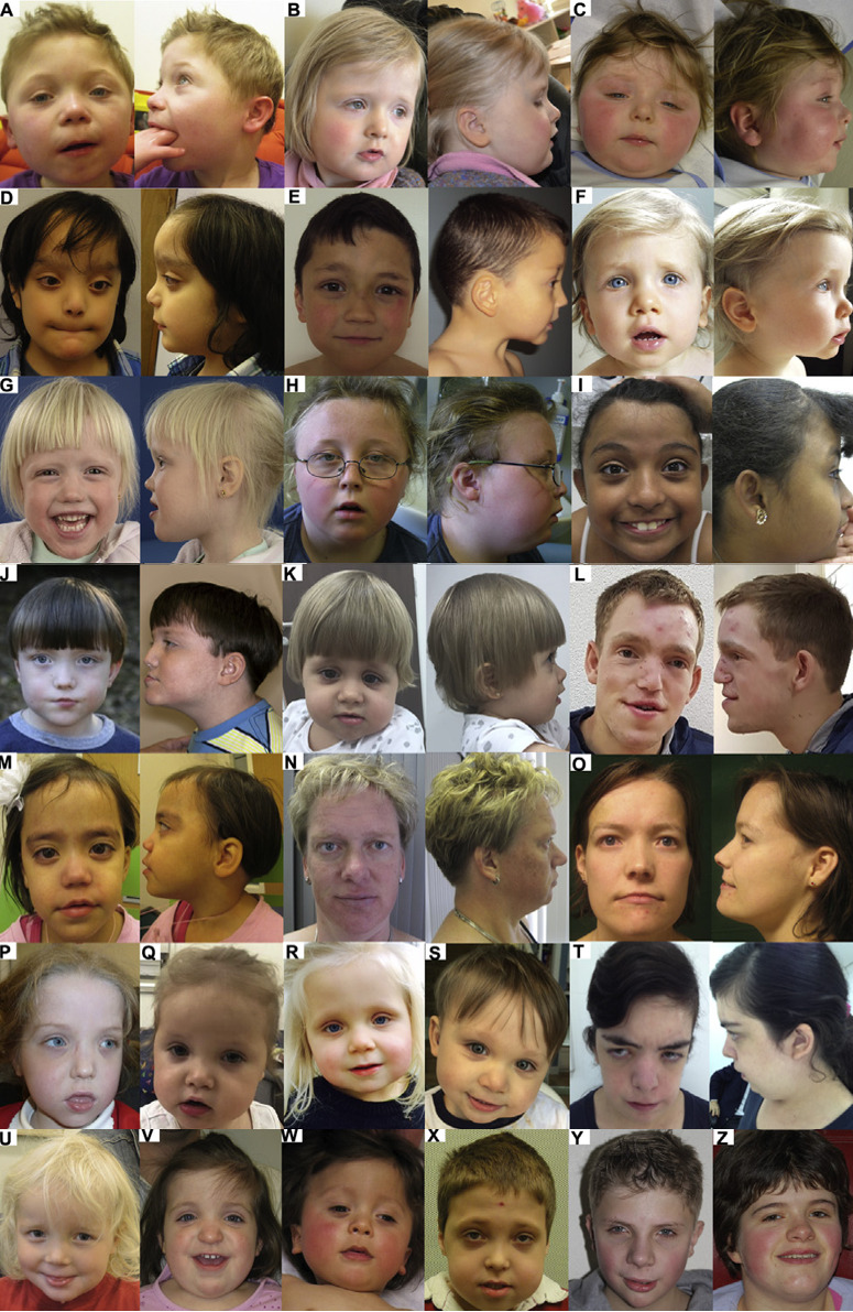
- Characteristic facial appearance including prominent forehead, high anterior hairline, downslanted palpebral fissures, prominent eyelashes, ear malformations, wide and depressed nasal bridge, short nose with full, upturned nasal tip, long philtrum, thin vermilion of the upper lip, pointed chin, and widely spaced teeth. See Figure 1.
- Feeding difficulties and gastrointestinal problems (e.g., gastroesophageal reflux disease, lack of satiation, frequent vomiting, constipation)
- Vison issues (e.g., strabismus, cortical visual impairment, hypermetropia) and various ophthalmologic defects
- Musculoskeletal anomalies (e.g., joint laxity, hand and foot anomalies, pectus deformities, plagiocephaly)
- Other features: endocrine manifestations, cardiac and renal anomalies, hearing loss, seizures, recurrent infections
MRI findings include atypical white matter lesions, wide ventricles, corpus callosum underdevelopment, cerebral atrophy, cortical dysplasia, and choroid cysts. Note that these findings are not sufficiently distinct to specifically suggest the diagnosis of ADNP-related disorder.
Establishing the Diagnosis
The diagnosis of ADNP-related disorder is established in a proband with suggestive findings and a heterozygous pathogenic (or likely pathogenic) variant in ADNP identified by molecular genetic testing (see Table 1).
Note: (1) Per ACMG/AMP variant interpretation guidelines, the terms "pathogenic variants" and "likely pathogenic variants" are synonymous in a clinical setting, meaning that both are considered diagnostic and both can be used for clinical decision making [Richards et al 2015]. Reference to "pathogenic variants" in this section is understood to include any likely pathogenic variants. (2) Identification of a heterozygous ADNP variant of uncertain significance does not establish or rule out the diagnosis.
Molecular genetic testing in a child with developmental delay or an older individual with intellectual disability typically begins with chromosomal microarray analysis (CMA). If CMA is not diagnostic, the next step is typically either a multigene panel or exome sequencing. Single-gene testing (sequence analysis of ADNP, followed by gene-targeted deletion/duplication analysis) may be indicated in individuals with the distinctive findings described in Suggestive Findings.
- Chromosomal microarray analysis (CMA) uses oligonucleotide or SNP array to detect genome-wide large deletions/duplications (including ADNP) that cannot be detected by sequence analysis.
- Single-gene testing. Sequence analysis of ADNP can be performed to detect small intragenic deletions/insertions and missense, nonsense, and splice site variants. Note: Depending on the sequencing method used, single-exon, multiexon, or whole-gene deletions/duplications may not be detected. If no variant is detected by the sequencing method used, the next step is to perform gene-targeted deletion/duplication analysis to detect exon and whole-gene deletions or duplications.
- A multigene panel that includes ADNP and other genes of interest (see Differential Diagnosis) may be considered to identify the genetic cause while limiting identification of variants of uncertain significance and pathogenic variants in genes that do not explain the underlying phenotype. Note: (1) The genes included in the panel and the diagnostic sensitivity of the testing used for each gene vary by laboratory and are likely to change over time. (2) Some multigene panels may include genes not associated with the condition discussed in this GeneReview (3) In some laboratories, panel options may include a custom laboratory-designed panel and/or custom phenotype-focused exome analysis that includes genes specified by the clinician. (4) Methods used in a panel may include sequence analysis, deletion/duplication analysis, and/or other non-sequencing-based tests.
- Comprehensive genomic testing does not require the clinician to determine which gene(s) are likely involved. Exome sequencing is most commonly used and yields results similar to a multigene panel with the additional advantage that exome sequencing includes genes recently identified as causing intellectual disability whereas some multigene panels may not. Genome sequencing is also possible.
- Epigenetic signature analysis / methylation array. Two distinctive epigenetic signatures (disorder-specific genome-wide changes in DNA methylation profiles) in peripheral blood leukocytes have been identified in individuals with ADNP-related disorder [Aref-Eshghi et al 2020]. Individuals with an ADNP pathogenic variant located outside the region between nucleotides 2000 and 2340 have a distinct epigenetic signature, whereas individuals with an ADNP pathogenic variant located between nucleotides 2000 and 2340 have a different epigenetic signature [Bend et al 2019, Breen et al 2020]. Epigenetic signature analysis of a peripheral blood sample or DNA banked from a blood sample can therefore be considered to clarify the diagnosis in individuals with clinical findings of ADNP-related disorder and a variant of uncertain clinical significance identified by molecular genetic testing. For an introduction to epigenetic signature analysis click here.
Table 1.
Molecular Genetic Testing Used in ADNP-Related Disorder
Clinical Characteristics
Clinical Description
The following clinical description is based on a published report of a large cohort of 78 individuals in whom a pathogenic variant in ADNP has been identified [Van Dijck et al 2019].
Of note, the oldest individual known to the authors to date is age 40 years [Author, personal observation].
Table 2.
Select Features of ADNP-Related Disorder
Development. Infants often have hypotonia. Developmental milestones are delayed: the average age to sit independently is 13 months, and the average walking age is 2.5 years. Speech impairment is prominent, with expressive language ranging from no words to sentences. Bladder training is delayed in half of affected individuals.
All have mild-to-severe intellectual disability.
Autism spectrum disorder (ASD), characterized by stereotypic behavior and impaired social interaction, is reported in 67% of the children. Children have a strong sensory interest (sensory processing disorder). A high pain threshold is common.
Additional behavior problems may include anxiety, obsessive compulsive disorder, aggressive behavior, temper tantrums, attention-deficient/hyperactivity disorder, and sleep problems.
Characteristic facial features include a prominent forehead, high anterior hairline, ptosis, downslanted palpebral fissures, prominent eyelashes, wide and depressed nasal bridge, short nose with full, upturned nasal tip, a long philtrum, thin vermilion of the upper lip, widely spaced teeth, pointed chin, and ear abnormalities including small low-set ears and protruding cup-shaped ears.
Gastrointestinal/feeding. Feeding difficulties and gastrointestinal problems are common, including decreased sucking or chewing, gastroesophageal reflux disease, lack of satiation, frequent vomiting, and constipation. Some individuals required a gastrostomy tube.
Vision issues. More than half have visual problems, most commonly hypermetropia or strabismus. Forty-one percent have cortical visual impairment. Ophthalmologic defects are diverse: ectropion, coloboma, congenital cataracts, nystagmus, everted or notched eyelid, or mild ptosis.
Hand abnormalities are present, including clinodactyly, polydactyly, small fifth fingers, fetal fingertip pads, prominent interphalangeal joints and distal phalanges, a single palmar crease, and nail anomalies.
Foot abnormalities include toe malformations, flat feet, and sandal gap.
Additional muscular skeletal features include joint laxity (38%), pectus deformities (22%) such as pectus excavatum, pectus carinatum or narrow thorax, or skull deformity (14%) including plagiocephaly, trigonocephaly, or brachycephaly.
Endocrine manifestations include thyroid hormone problems (15%, mainly hypothyroidism) and/or growth hormone deficiency. Some have signs of early pubertal development.
Growth. Birth weight, length, and occipitofrontal circumference are within the normal range. Several develop truncal obesity. Twenty-three percent of the individuals have short stature (height <-2 SD in individuals reported with age range of 2-23 years). Growth hormone deficiency is present in 11%.
Cardiac anomalies. Atrial septal defect is the most common. Less frequent cardiac defects include: patent ductus arteriosus, patent foramen ovale, mitral valve prolapse, ventricular septal defect, and other cardiovascular malformations.
Seizures. Some children have seizures (16%) including absence seizures, focal seizures with reduced awareness, and epilepsy with continuous spike and waves during slow-wave sleep.
Renal. Renal anomalies include narrow ureters or bilateral vesicoureteral reflux.
Recurrent infections. Most children have recurrent infections, including upper respiratory and urinary tract infections (51%).
Other
- Submucous cleft palate (1 individual)
- Metopic craniosynostosis (2 individuals)
Genotype-Phenotype Correlations
There is no evidence for a clinically relevant impact of a specific pathogenic variant on the clinical presentation. However, it was noticed that individuals with a p.Tyr719Ter pathogenic variant walked later and had a higher pain threshold than those with other ADNP pathogenic variants [Van Dijck et al 2019].
Penetrance
Penetrance is less than 100%; In two families reported to date, probands diagnosed with ADNP-related disorder inherited a pathogenic variant from an unaffected parent [Van Dijck et al 2019].
Prevalence
The prevalence of pathogenic variants in ADNP is estimated at 0.17% of individuals with ASD (95% binomial confidence interval: 0.083%-0.32%). It is one of the most common known single-gene causes of ASD [Helsmoortel et al 2014].
Genetically Related (Allelic) Disorders
No phenotypes other than those discussed in this GeneReview are known to be associated with germline pathogenic variants in ADNP.
Differential Diagnosis
Phenotypic features associated with ADNP pathogenic variants are not sufficient to diagnose ADNP-related disorder. All genes known to be associated with intellectual disability * should be included in the differential diagnosis of ADNP-related disorder.
* More than 200 have been identified; see OMIM Autosomal Dominant, Autosomal Recessive, Nonsyndromic X-Linked, and Syndromic X-Linked Intellectual Developmental Disorder Phenotypic Series.
Management
Evaluations Following Initial Diagnosis
To establish the extent of disease and needs in an individual diagnosed with ADNP-related disorder, the evaluations summarized in Table 3 (if not performed as part of the evaluation that led to the diagnosis) are recommended.
Table 3.
Recommended Evaluations Following Initial Diagnosis in Individuals with ADNP-Related Disorder
Treatment of Manifestations
Treatment is symptomatic; no specific therapy is available. Routine medical care by a pediatrician or primary care physician is recommended.
Table 4.
Treatment of Manifestations in Individuals with ADNP-Related Disorder
Surveillance
Table 5.
Recommended Surveillance for Individuals with ADNP-Related Disorder
Evaluation of Relatives at Risk
See Genetic Counseling for issues related to testing of at-risk relatives for genetic counseling purposes.
Therapies Under Investigation
Administration of NAP, a neuroprotective octapeptide (NAPVSPIQ), has been reported to ameliorate some of the cognitive abnormalities observed in a knockout mouse model [Bassan et al 1999, Vulih-Shultzman et al 2007]. It restores learning and memory and reduces neurodegeneration in Adnp+/− mice. The drug name for NAP is davunetide, a candidate for treatment of multiple selected neurologic disorders. Intranasal and intravenous formulations of the drug have been shown to cross the blood-brain barrier. Phase II and Phase III clinical trials showed good tolerance without significant side effects. Although it is not known whether the disease is the result of a loss of function of ADNP and although the knockout mouse model has not been evaluated for autistic traits, the observations raise hope for treatment in individuals with ADNP-related disorder [Vandeweyer et al 2014].
A Phase II trial with the drug ketamine in children with ADNP-related disorder is currently recruiting.
Search ClinicalTrials.gov in the US and EU Clinical Trials Register in Europe for access to information on clinical studies for a wide range of diseases and conditions.
Genetic Counseling
Genetic counseling is the process of providing individuals and families with information on the nature, mode(s) of inheritance, and implications of genetic disorders to help them make informed medical and personal decisions. The following section deals with genetic risk assessment and the use of family history and genetic testing to clarify genetic status for family members; it is not meant to address all personal, cultural, or ethical issues that may arise or to substitute for consultation with a genetics professional. —ED.
Mode of Inheritance
ADNP-related disorder is an autosomal dominant disorder typically caused by a de novo pathogenic variant.
Risk to Family Members
Parents of a proband
- Most probands reported to date with ADNP-related disorder whose parents have undergone molecular genetic testing have the disorder as the result of a de novo ADNP pathogenic variant.
- In two families reported to date, probands diagnosed with ADNP-related disorder inherited a pathogenic variant from an unaffected parent [Van Dijck et al 2019].
- Molecular genetic testing is recommended for the parents of the proband to confirm their genetic status and to allow reliable recurrence risk counseling.
- If the pathogenic variant identified in the proband is not identified in either parent, the following possibilities should be considered:
- The proband has a de novo pathogenic variant. Note: A pathogenic variant is reported as "de novo" if: (1) the pathogenic variant found in the proband is not detected in parental DNA; and (2) parental identity testing has confirmed biological maternity and paternity. If parental identity testing is not performed, the variant is reported as "assumed de novo" [Richards et al 2015].
- The proband inherited a pathogenic variant from a parent with germline (or somatic and germline) mosaicism. Note: Testing of parental leukocyte DNA may not detect all instances of somatic mosaicism.
Sibs of a proband. The risk to the sibs of the proband depends on the genetic status of the proband's parents:
- If the ADNP pathogenic variant found in the proband cannot be detected in the leukocyte DNA of either parent, the recurrence risk to sibs is estimated to be 1% because of the theoretic possibility of parental germline mosaicism [Rahbari et al 2016].
- If a parent of the proband is known to have the ADNP pathogenic variant identified in the proband, the risk to the sibs of inheriting the variant is 50%.
Offspring of a proband
- Each child of an individual with ADNP-related disorder has a 50% chance of inheriting the ADNP pathogenic variant.
- Individuals with ADNP-related syndromic autism are not known to reproduce.
Other family members
- The risk to other family members depends on the genetic status of the proband's parents: if a parent has the ADNP pathogenic variant, the parent's family members may be at risk.
- Given that most probands with ADNP-related disorder reported to date have the disorder as a result of a de novo ADNP pathogenic variant, the risk to other family members is presumed to be low.
Related Genetic Counseling Issues
Family planning
- The optimal time for determination of genetic risk and discussion of the availability of prenatal/preimplantation genetic testing is before pregnancy.
- It is appropriate to offer genetic counseling (including discussion of potential risks to offspring and reproductive options) to the parents of an affected individual.
Prenatal Testing and Preimplantation Genetic Testing
Once the ADNP pathogenic variant has been identified in an affected family member, prenatal and preimplantation genetic testing are possible.
Differences in perspective may exist among medical professionals and within families regarding the use of prenatal testing. While most centers would consider use of prenatal testing to be a personal decision, discussion of these issues may be helpful.
Resources
GeneReviews staff has selected the following disease-specific and/or umbrella support organizations and/or registries for the benefit of individuals with this disorder and their families. GeneReviews is not responsible for the information provided by other organizations. For information on selection criteria, click here.
- ADNP Kids
- ADNP Kids Research FoundationEmail: ADMIN@adnpfoundation.org
- ADNP Kids-HVDAS BelgiumBelgiumEmail: adnpkids.hvdasbelgium@gmail.com
- Amis de ADNP FranceFranceEmail: caroleherman2012@gmail.com
- American Association on Intellectual and Developmental Disabilities (AAIDD)Phone: 202-387-1968
- CDC - Child DevelopmentPhone: 800-232-4636
- MedlinePlus
- Simons Searchlight RegistrySimons Searchlight aims to further the understanding of rare genetic neurodevelopmental disorders.Phone: 855-329-5638Fax: 570-214-7327Email: coordinator@simonssearchlight.org
Molecular Genetics
Information in the Molecular Genetics and OMIM tables may differ from that elsewhere in the GeneReview: tables may contain more recent information. —ED.
Table A.
ADNP-Related Disorder: Genes and Databases
Table B.
OMIM Entries for ADNP-Related Disorder (View All in OMIM)
Molecular Pathogenesis
ADNP contains five exons, the last three of which are translated. The protein contains nine zinc fingers and three other functional domains, including NAP, an 8-amino-acid neuroprotectant peptide (NAPVSIPQ), a DNA-binding homeobox domain, and a HP1-binding motif.
ADNP is a vasoactive intestinal peptide (VIP)-responsive gene. VIP, a neuroprotective peptide, is active during embryonic development, in particular during the time of neuronal tube closure. It protects damaged nerve cells from cell death by inducing glia-derived, survival-promoting substances [Helsmoortel et al 2014].
Wild type ADNP directly binds genomic DNA and mediates the recruitment of the BAF complex through its C-terminal end. The C-terminal end is truncated in all individuals with an ADNP-related disorder. It has been hypothesized that the mutated protein still binds to the DNA, but is no longer capable of recruiting the BAF complex, leading to diminished functionality of the complex as a whole and ultimately to deregulation of several cellular processes [Helsmoortel et al 2014]. ADNP has also been shown to interact with the chromatin-remodeling gene CHD4 [Ostapcuk et al 2018].
Mechanism of disease causation. The mutational mechanism is not known, but it has been hypothesized that the mutated ADNP protein competes with the wild type protein to bind with the BAF complexes (the functional eukaryotic equivalent of the SWI/SNF complex that is involved in the regulation of gene expression in yeast) [Helsmoortel et al 2014].
Table 6.
Notable ADNP Pathogenic Variants
Chapter Notes
Author Notes
The research group Cognitive Genetics is part of the research cluster Medical Genetics of the Faculty of Pharmaceutical, Biomedical, and Veterinary Sciences of the University of Antwerp. Our mission is to identify genetic causes of cognitive disorders and to study the molecular defects in order to eventually develop rational therapy.
Acknowledgments
We thank the families with individuals affected by an ADNP pathogenic variant who are participating in our research programs.
Author History
Céline Helsmoortel, MSc; University of Antwerp (2016-2021)
Frank Kooy, PhD (2016-present)
Anke Van Dijck, MD, PhD (2016-present)
Geert Vandeweyer, PhD (2016-present)
Revision History
- 6 October 2022 (sw) Revision: epigenetic signature analysis (Establishing the Diagnosis)
- 15 April 2021 (sw) Comprehensive update posted live
- 7 April 2016 (bp) Review posted live
- 18 December 2015 (avd) Original submission
References
Literature Cited
- Aref-Eshghi E, Kerkhof J, Pedro VP, Groupe DI. France, Barat-Houari M, Ruiz-Pallares N, Andrau JC, Lacombe D, Van-Gils J, Fergelot P, Dubourg C, Cormier-Daire V, Rondeau S, Lecoquierre F, Saugier-Veber P, Nicolas G, Lesca G, Chatron N, Sanlaville D, Vitobello A, Faivre L, Thauvin-Robinet C, Laumonnier F, Raynaud M, Alders M, Mannens M, Henneman P, Hennekam RC, Velasco G, Francastel C, Ulveling D, Ciolfi A, Pizzi S, Tartaglia M, Heide S, Héron D, Mignot C, Keren B, Whalen S, Afenjar A, Bienvenu T, Campeau PM, Rousseau J, Levy MA, Brick L, Kozenko M, Balci TB, Siu VM, Stuart A, Kadour M, Masters J, Takano K, Kleefstra T, de Leeuw N, Field M, Shaw M, Gecz J, Ainsworth PJ, Lin H, Rodenhiser DI, Friez MJ, Tedder M, Lee JA, DuPont BR, Stevenson RE, Skinner SA, Schwartz CE, Genevieve D, Sadikovic B. Evaluation of DNA methylation episignatures for diagnosis and phenotype correlations in 42 mendelian neurodevelopmental disorders. Am J Hum Genet. 2020;106:356–70. [PMC free article: PMC7058829] [PubMed: 32109418]
- Bassan M, Zamostiano R, Davidson A, Pinhasov A, Giladi E, Perl O, Bassan H, Blat C, Gibney G, Glazner G, Brenneman DE, Gozes I. Complete sequence of a novel protein containing a femtomolar-activity-dependent neuroprotective peptide. J Neurochem. 1999;72:1283–93. [PubMed: 10037502]
- Bend EG, Aref-Eshghi E, Everman DB, Rogers RC, Cathey SS, Prijoles EJ, Lyons MJ, Davis H, Clarkson K, Gripp KW, Li D, Bhoj E, Zackai E, Mark P, Hakonarson H, Demmer LA, Levy MA, Kerkhof J, Stuart A, Rodenhiser D, Friez MJ, Stevenson RE, Schwartz CE, Sadikovic B. Gene domain-specific DNA methylation episignatures highlight distinct molecular entities of ADNP syndrome. Clin Epigenetics. 2019;11:64. [PMC free article: PMC6487024] [PubMed: 31029150]
- Breen MS, Garg P, Tang L, Mendonca D, Levy T, Barbosa M, Arnett AB, Kurtz-Nelson E, Agolini E, Battaglia A, Chiocchetti AG, Freitag CM, Garcia-Alcon A, Grammatico P, Hertz-Picciotto I, Ludena-Rodriguez Y, Moreno C, Novelli A, Parellada M, Pascolini G, Tassone F, Grice DE, Di Marino D, Bernier RA, Kolevzon A, Sharp AJ, Buxbaum JD, Siper PM, De Rubeis S. Episignatures stratifying Helsmoortel-Van Der Aa syndrome show modest correlation with phenotype. Am J Hum Genet. 2020;107:555–63. [PMC free article: PMC7477006] [PubMed: 32758449]
- Coe BP, Witherspoon K, Rosenfeld JA, van Bon BW, Vulto-van Silfhout AT, Bosco P, Friend KL, Baker C, Buono S, Vissers LE, Schuurs-Hoeijmakers JH, Hoischen A, Pfundt R, Krumm N, Carvill GL, Li D, Amaral D, Brown N, Lockhart PJ, Scheffer IE, Alberti A, Shaw M, Pettinato R, Tervo R, de Leeuw N, Reijnders MR, Torchia BS, Peeters H, O'Roak BJ, Fichera M, Hehir-Kwa JY, Shendure J, Mefford HC, Haan E, Gécz J, de Vries BB, Romano C, Eichler EE. Refining analyses of copy number variation identifies specific genes associated with developmental delay. Nat Genet. 2014;46:1063–71. [PMC free article: PMC4177294] [PubMed: 25217958]
- De Rubeis S, He X, Goldberg AP, Poultney CS, Samocha K, Cicek AE, Kou Y, Liu L, Fromer M, Walker S, Singh T, Klei L, Kosmicki J, Shih-Chen F, Aleksic B, Biscaldi M, Bolton PF, Brownfeld JM, Cai J, Campbell NG, Carracedo A, Chahrour MH, Chiocchetti AG, Coon H, Crawford EL, Curran SR, Dawson G, Duketis E, Fernandez BA, Gallagher L, Geller E, Guter SJ, Hill RS, Ionita-Laza J, Jimenz Gonzalez P, Kilpinen H, Klauck SM, Kolevzon A, Lee I, Lei I, Lei J, Lehtimäki T, Lin CF, Ma'ayan A, Marshall CR, McInnes AL, Neale B, Owen MJ, Ozaki N, Parellada M, Parr JR, Purcell S, Puura K, Rajagopalan D, Rehnström K, Reichenberg A, Sabo A, Sachse M, Sanders SJ, Schafer C, Schulte-Rüther M, Skuse D, Stevens C, Szatmari P, Tammimies K, Valladares O, Voran A, Li-San W, Weiss LA, Willsey AJ, Yu TW, Yuen RK, Cook EH, Freitag CM, Gill M, Hultman CM, Lehner T, Palotie A, Schellenberg GD, Sklar P, State MW, Sutcliffe JS, Walsh CA, Scherer SW, Zwick ME, Barett JC, Cutler DJ, Roeder K, Devlin B, Daly MJ, Buxbaum JD, et al. Synaptic, transcriptional and chromatin genes disrupted in autism. Nature. 2014;515:209–15. [PMC free article: PMC4402723] [PubMed: 25363760]
- Deciphering Developmental Disorders Study Group. Large-scale discovery of novel genetic causes of developmental disorders. Nature. 2015;519:223–8. [PMC free article: PMC5955210] [PubMed: 25533962]
- Helsmoortel C, Vulto-van Silfhout AT, Coe BP, Vandeweyer G, Rooms L, van den Ende J, Schuurs-Hoeijmakers JH, Marcelis CL, Willemsen MH, Vissers LE, Yntema HG, Bakshi M, Wilson M, Witherspoon KT, Malmgren H, Nordgren A, Annerén G, Fichera M, Bosco P, Romano C, de Vries BB, Kleefstra T, Kooy RF, Eichler EE, Van der Aa N. A. SWI/SNF-related autism syndrome caused by de novo mutations in ADNP. Nat Genet. 2014;46:380–4. [PMC free article: PMC3990853] [PubMed: 24531329]
- Huynh MT, Boudry-Labis E, Massard A, Thuillier C, Delobel B, Duban-Bedu B, Vincent-Delorme C. A heterozygous microdeletion of 20q13.13 encompassing ADNP gene in a child with Helsmoortel-van der Aa syndrome. Eur J Hum Genet. 2018;26:1497–501. [PMC free article: PMC6138634] [PubMed: 29899371]
- Ostapcuk V, Mohn F, Carl SH, Basters A, Hess D, Iesmantavicius V, Lampersberger L, Flemr M, Pandey A, Thomä NH, Betschinger J, Bühler M. Activity-dependent neuroprotective protein recruits HP1 and CHD4 to control lineage-specifying genes. Nature. 2018;557:739–43. [PubMed: 29795351]
- Pescosolido MF, Schwede M, Johnson Harrison A, Schmidt M, Gamsiz ED, Chen WS, Donahue JP, Shur N, Jerskey BA, Phornphutkul C, Morrow EM. Expansion of the clinical phenotype associated with mutations in activity-dependent neuroprotective protein. J Med Genet. 2014;51:587–9. [PMC free article: PMC4135390] [PubMed: 25057125]
- Rahbari R, Wuster A, Lindsay SJ, Hardwick RJ, Alexandrov LB, Turki SA, Dominiczak A, Morris A, Porteous D, Smith B, Stratton MR, Hurles ME, et al. Timing, rates and spectra of human germline mutation. Nat Genet. 2016;48:126–33. [PMC free article: PMC4731925] [PubMed: 26656846]
- Richards S, Aziz N, Bale S, Bick D, Das S, Gastier-Foster J, Grody WW, Hegde M, Lyon E, Spector E, Voelkerding K, Rehm HL, et al. Standards and guidelines for the interpretation of sequence variants: a joint consensus recommendation of the American College of Medical Genetics and Genomics and the Association for Molecular Pathology. Genet Med. 2015;17:405–24. [PMC free article: PMC4544753] [PubMed: 25741868]
- Stenson PD, Mort M, Ball EV, Chapman M, Evans K, Azevedo L, Hayden M, Heywood S, Millar DS, Phillips AD, Cooper DN. The Human Gene Mutation Database (HGMD®): optimizing its use in a clinical diagnostic or research setting. Hum Genet. 2020;139:1197–207. [PMC free article: PMC7497289] [PubMed: 32596782]
- Van Dijck A, Vulto-van Silfhout AT, Cappuyns E, van der Werf IM, Mancini GM, Tzschach A, Bernier R, Gozes I, Eichler EE, Romano C, Lindstrand A, Nordgren A., ADNP Consortium. Kvarnung M, Kleefstra T, de Vries BB, Küry S, Rosenfeld JA, Meuwissen ME, Vandeweyer G, Kooy RF. Clinical presentation of a complex neurodevelopmental disorder caused by mutations in ADNP. Biol Psychiatry. 2019;85:287–97. [PMC free article: PMC6139063] [PubMed: 29724491]
- Vandeweyer G, Helsmoortel C, Van Dijck A, Vulto-van Silfhout AT, Coe BP, Bernier R, Gerdts J, Rooms L, van den Ende J, Bakshi M, Wilson M, Nordgren A, Hendon LG, Abdulrahman OA, Romano C, de Vries BB, Kleefstra T, Eichler EE, Van der Aa N, Kooy RF. The transcriptional regulator ADNP links the BAF (SWI/SNF) complexes with autism. Am J Med Genet C Semin Med Genet. 2014;166C:315–26. [PMC free article: PMC4195434] [PubMed: 25169753]
- Vulih-Shultzman I, Pinhasov A, Mandel S, Grigoriadis N, Touloumi O, Pittel Z, Gozes I. Activity-dependent neuroprotective protein snippet NAP reduces tau hyperphosphorylation and enhances learning in a novel transgenic mouse model. J Pharmacol Exp Ther. 2007;323:438–49. [PubMed: 17720885]
Publication Details
Author Information and Affiliations
De Mick, Klina Hospital Brasschaat
Brasschaat, Belgium
Edegem, Belgium
Antwerp University Hospital
Edegem, Belgium
Department of Medical Genetics
University of Antwerp
Edegem, Belgium
Publication History
Initial Posting: April 7, 2016; Last Revision: October 6, 2022.
Copyright
GeneReviews® chapters are owned by the University of Washington. Permission is hereby granted to reproduce, distribute, and translate copies of content materials for noncommercial research purposes only, provided that (i) credit for source (http://www.genereviews.org/) and copyright (© 1993-2024 University of Washington) are included with each copy; (ii) a link to the original material is provided whenever the material is published elsewhere on the Web; and (iii) reproducers, distributors, and/or translators comply with the GeneReviews® Copyright Notice and Usage Disclaimer. No further modifications are allowed. For clarity, excerpts of GeneReviews chapters for use in lab reports and clinic notes are a permitted use.
For more information, see the GeneReviews® Copyright Notice and Usage Disclaimer.
For questions regarding permissions or whether a specified use is allowed, contact: ude.wu@tssamda.
Publisher
University of Washington, Seattle, Seattle (WA)
NLM Citation
Van Dijck A, Vandeweyer G, Kooy F. ADNP-Related Disorder. 2016 Apr 7 [Updated 2022 Oct 6]. In: Adam MP, Feldman J, Mirzaa GM, et al., editors. GeneReviews® [Internet]. Seattle (WA): University of Washington, Seattle; 1993-2024.

